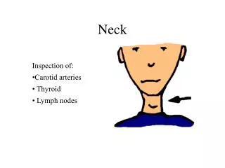
Neck
Neck . 山东大学医学院 解剖教研室 李振华. The muscles of neck. Superficial group Platysma 颈阔肌 , a thin sheet-like muscle of facial expression Sternocleidomastoid 胸锁乳突肌. Suprahyoid muscles Digastric 二腹肌 Stylohyoid 茎突舌骨肌 Mylohyoid 下颌舌骨肌 Geniohyoid 颏舌骨肌


Neck
E N D
Presentation Transcript
Neck 山东大学医学院 解剖教研室 李振华
The muscles of neck • Superficial group • Platysma 颈阔肌,a thin sheet-like muscle of facial expression • Sternocleidomastoid 胸锁乳突肌
Suprahyoid muscles • Digastric 二腹肌 • Stylohyoid 茎突舌骨肌 • Mylohyoid 下颌舌骨肌 • Geniohyoid 颏舌骨肌 Elevate (raise) hyoid bone and depress mandible.
Infrahyoid muscle • Sternohyoid 胸骨舌骨肌 • Sternothyroid 胸骨甲状肌 • Thyrohyoid甲状舌骨肌 • Omohyoid 肩胛舌骨肌 Depress hyoid or larynx after elevation
Deep group • Lateral • Scalenus anterior 前斜角肌 • Scalenus medius 中斜角肌 • Scalenus posterior 后斜角肌 • Medial • longus capitis 头长肌 • longus colli 颈长肌 Flex the head, bends the neck forward
Major muscles of the neck Sternocleidomastoid • Origin: manubrium and sternal end of clavicle • Insertion: mastoid process of temporal bone • Action: contraction of one muscle draws head toward the same side, and turn face to opposite side; both muscles act together to draw head backward
Scalenus anterior • Origin: transverse processes of C3-C6. • Insertion: tubercle for scalenus anterior • Action: unilateral, bends neck laterally; bilateral, elevate first rib, an accessory muscle of inspiration; if rib is fixed, flex neck anteriorly Scalene fissure 斜角肌间隙Above the first rib, there is a triangular space between scalenus anterior and midius. The brachial plexus and the subclavine a. emerge from this space.
The arteries of neck Common carotid artery颈总动脉 • Origin (arises from) • Brachiocephalic trunk on the right • Aortic arch on the left • Ascends in neck to upper border of thyroid cartilage; bifurcates into internal and external carotid arteries
Carotid sinus颈动脉窦(baroreceptor), located at a localizes dilation of terminal part of common carotid artery or beginning of internal carotid artery, sensitive to blood pressure changes • Carotid glomus 颈动脉小球(chemoreceptor), lies posterior to the point of bifurcation of common carotid artery, senses changes in blood carbon dioxide (oxygen) levels
Branches of external carotid a. • Superior thyroid a. 甲状腺上动脉-descends to supply upper pole of thyroid gland and larynx • Lingual a. 舌动脉 • Facial a. 面动脉 • Occipital a.枕动脉 • Posterior auricular a. 耳后动脉 • Maxillary a. 上颌动脉 • Superficial temporal a. 颞浅动脉
Subclavian artery 锁骨下动脉 • Origin (arises from) • Brachiocephalic trunk on right • Aortic arch on left • Becomes axillary artery at lateral border of first rib • Branches • Vertebral a. 椎动脉 • Internal thoracic a. 胸廓内动脉 • Thyrocervical trunk甲状颈干 • Inferior thyroid artery 甲状腺下动脉-supplies inferior pole of thyroid gland • Costocervical trunk 肋颈干
Veins Draining the neck Internal jugular vein颈内静脉 • Begin at jugular foramen, descending to join the subclavian vein to form brachiocephalic vein • Lies lateral first to internal and then to common carotid a. within carotid sheath • Chief extracranial tributaries • Common facial vein 面总静脉 • Lingual v. 舌静脉 • Pharyngeal v. 咽静脉 • Superior thyroid v.甲状腺上静脉 • Middle thyroid v. 甲状腺中静脉
Subclavian vein • It is an continuation of axillary vein at the lateral border of first rib • Joins internal jugular vein to form the brachiocephalic vein. Angle of union is termed venous angle静脉角
External jugular vein颈外静脉 • Formed behind angle of mandible by union of posterior auricular, posterior branch of retromandibular and occipital vein • Crossing sternocleidomastoid to enter subclavian vein Anterior jugular vein 颈前静脉 • Drains submandibular and anterior neck regions • Descends near midline, runs posterior to sternal end of sternocleidomastoid to drain into external jugular vein or subclavian vein
Lymph nodes of neck Anterior cervical lymph nodes • Superficial anterior cervical lymph nodes • Deep anterior cervical lymph nodes Lateral cervical lymph node • Superficial lateral cervical lymph nodes-lie along the external jugular vein
Deep lateral cervical lymph nodes • Extend along the internal jugular vein from the base of skull to the root of neck, divided into superior deep lateral cervical lymph nodes and inferior deep lateral cervical lymph nodes • Receive lymphatic vessels from head, neck, tongue, larynx, cervical parts of esophagus and trachea, thyroid gland, upper parts of the thoracic wall and breast • Efferent vessels form the jugular trunk-the left jugular trunk joins the thoracic duct and right may joint the right lymphatic duct
Superior deep lateral cervical lymph nodes 颈外侧上深淋巴结 • Jugulodigastric lymph node 颈内静脉二腹肌淋巴结: lies at the junction of posterior belly of digastric and internal jugular vein Inferior deep lateral cervical lymph nodes 颈外侧下深淋巴结 • Juguloomohyoid lymph node颈内静脉肩胛舌骨肌淋巴结: lies at the junction of the intermediate tendon of omohyoid and internal jugular vein • Supraclavicular lymph nodes锁骨上淋巴结: lie along the subclavian artery
Right lymphatic duct 右淋巴导管 • Formed by union of right jugular, subclavian, and bronchomediastinal trunks • Ends by entering the right venous angle • Receives lymph from right half of head, neck, thorax and right upper limb Thoracic duct 胸导管 • At the roof of the neck, it turns laterally and arches forwards and descends to enter the left venous angle • Just before termination, it receives the left jugular, subclavian and bronchomediastinal trunks
Cervical plexus Formation: formed by anterior rami of C1-C4 spinal nerves Position: lies in front of the origin of levator scapulae and scalenus medius and deep to the superior part of the sternocleidomastoid
Branches • Cutaneous branches: emerge around middle of posterior border of sternocleidomastoid, to supply skin of neck and scalp between auricle and external occipital protuberance • Lesser occipital n. 枕小神经 • Greet auricular n. 耳大神经 • Transverse nerve of neck 颈横神经 • Supraclavicular n. 锁骨上神经
Muscular branches: supply the deep muscles of neck • Phrenic nerve膈神经(anterior rami of C3-C5) • Lies on anterior scalene, deep to fascia • To diaphragm (motor and sensory) • Ansa cervicalis 颈袢: • Hypoglossal nerve gives off superior root of ansa (descendens hypoglossi), composed of fibers picked up from nerve C1 • Joins inferior root of ansa (descendens cervicalis, C2and C3) to form a loop, the ansa cervicalis, which supplies infrahyoid muscles
Vagus nerve (Ⅹ) • Leaves skull via jugular foramen • Descends in the neck in carotid sheath between internal (or common) carotid artery and internal jugular vein
Branches • Superior laryngeal nerve 喉上神经passes down side of pharynx and given rise to • Internal branch 内支which pierces thyrohyoid membrane to innervates mucous membrane of larynx above fissure of glottis • External branch外支 which innervates cricothyroid • Cervical cardiac branches 颈心支: descending to terminate in cardiac plexus • Recurrent laryngeal nerves喉返神经 • Ascend in tracheo-esophageal groove • Enter larynx posterior to cricothyroid joint, the nerve is now called inferior laryngeal nerve • Innervations: laryngeal mucosa below fissure of glottis , all laryngeal laryngeal muscles except cricothyroid
Accessory n. (Ⅺ ) 副神经 • Deep to posterior belly of digastric • Supplies sternoclidomastoid and trapezius muscle Hypoglossal n.(Ⅻ) 舌下神经 • Descends between internal carotid a. and internal jugular v., hooks around external carotid a., to lie on the hyoglossus before entering tongue • Supplies muscles of tongue
Cervical part of sympathetic trunk 颈交感干 • Formed by superior , middle and inferior cervical ganglia and interganglionic branches • Superior cervical ganglion: largest, situated in front of transverse processes of C1~C3 vertebra • Middle cervical ganglion: smallest, is at level of transverse processes of C6 vertebra • Inferior cervical ganglion: situated at level of C7 vertebra, and may be fused with first thoracic ganglion to form cervicothoracic ganglion 颈胸神经节
Regional anatomy of neck 山东大学医学院 解剖教研室 李振华
Parts and regions of the neck Boundaries • Superior-inferior border of mandible, angle of mandible, tip of mastoid process, superior nuchal line, external occipital protuberance • Inferior-jugular notch, sternoclavicular joint, superior border of clavicle, acromion, spinous processes of C7
Landmarks • Hyoid bone • Thyroid cartilage • Cricoid cartilage • Catotid tubercle • Sternocleidomastoid • Suprasternal fossa • Greater supraclaviclar fossa
Regions • Neck 颈 • Anterior region of neck • Lateral region of neck • Sternocleidomastoid region • Nape 项部
Anterior region of neck • Suprahyoid region 舌骨上区 • Submental triangle 颏下三角 • Submandibular triangle 下颌下三角 • Infrahyoid region 舌骨下区 • Carotid triangle 颈动脉三角 • Muscular triangle 肌三角
Lateral region of neck • Occipital triangle 枕三角 • supraclavicular triangle 锁骨上三角 (greatsupraclavicular fossa) 锁骨上大窝
Skin • The natural line of cleavage of the skin are constant and run almost horizontally around the neck
Superficial fascia • Consists of fat and connective tissue • Contents • Platysma • Superficial veins • Anterior jugular v. • External jugular v. • Cutaneous nerves • Lesser occipital n. • Greet auricular n. • Transverse nerve of neck • Supraclavicular n. • Cervical branch of facial n.
Cervical fascia颈筋膜-three layers Superficial layer (investing fascia 封套筋膜) • Encloses trapezius, sternocleidomastoid, posterior belly of digastric and parotid and submandibular glands • Attached to bony landmarks of upper and lower boundaries of neck and zygomatic arch of face
Prevertebral layer气管前层 • Encloses viscera of neck: pharynx, larynx, trachea, esophagus, thyroid gland and parathyroid glands • Completely surrounds thyroid gland, forming a sheet for it, and bind the gland to larynx • Extends from hyoid bone to fibrous pericardium of superior mediastinum
Prevertebral layer 椎前层 • Lies anterior to bodies of cervical vertebrae and prevertebral muscles; extends from base of skull downward into the superior mediastinum, continuous with anterior longitudinal lig. and endothoracic fascia • Covers subclavian vessels and roots of brachial plexus • Extends into upper limb as axillary sheath
Carotid sheath 颈动脉鞘 • Formed by components of all three layers of deep cervical fascia • Contains common and internal carotid arteries, internal jugular vein, and vagus nerve • Ansa cervicalis located on its anterior surface
Suprasternal space 胸骨上间隙 • Over the lower part of the anterior triangle, the inversting fascia splits into two layers, which are attached to the anterior and posterior margins of the upper border of the manubrium, between these two layers is a slitlike space, called the suprasternal space • Contains connective tissue, and sometimes a lymph node
Pretracheal space 气管前间隙 • Lies between pretracheal layer and cervical part of trachea • Contains arteria thyroidea ima, inferior thyroid v., unpaired thyroid venous plexus, brachiocephalic trunk and left brachiocephalic v.
Retropharyngeal space咽后间隙 • Lies between prevertebral layer and buccopharyngeal fascia Prevertebral space椎前间隙 • Lies between prevertebral muscles, cervical part of vertebral column and prevertebral layer
Suprahyoid region Submental triangle 颏下三角 • Lies below the chin and is bounded laterally by anterior bellies of digastric, and inferiorly by the body of hyoid bone • Covered by skin, superficial fascia and investing fascia • Floor-mylohyoid muscles • Contents-submental lymph nodes
Submandibular triangle 下颌下三角 • Bounded by anterior and posterior bellies of digastric and lower border of the body of the mandible • Covered by skin, superficial fascia, platysma and investing fascia • Floor- mylohyoid, hyoglossus and middle constrictor of pharynx • Contents-submandibular gland, facial a., v., hypoglossal n. and v., lingual n., submandibular ganglion and submandibular lymph nodes
Infrahyoid region Carotid triangle颈动脉三角 • Bounded by anterior border of sternocleidomastoid, superior belly of omohyoid and posterior belly of digastic muscles • Covered by skin, superficial fascia, platysma and investing fascia • Floor-prevertebral fascia and lateral wall of pharynx • Contents-common carotid a. and its branches, internal jugular v. and its tributaries, hypoglossal n. with its descending branches, the accessory and vagus nerves, and part of the chain of deep cervical lymph nodes
Posterior belly of digastic muscle • Superficial-great auricular n., retromandibular v., cervical branch of facial n. • Deep-internal and external carotid a., internal jugular v., Ⅹ~Ⅻ cranial n. and cervical part of sympathetic trunk • Superior-posterior auricular a., facial a. and glossopharyngeal n. • Infeior-occipital a. and hypoglossal n.
Muscular triangle肌三角 • Bounded by midline of the neck, superior belly of the omohyoid and anterior border of the sternocleidomastoid. • Covered by skin, superficial fascia, platysma, anterior jugular v., coutaneous n. and investing fascia • Floor-prevertebral fascia • Contents-sternohyoid, superior belly of omohyoid, sternothyroid, thyrohyoid, thyroid gland, parathyroid gland, cervical part of trachea and esophagus
Thyroid gland 甲状腺 Shape and position • H-shape • Left and right lobes: lie on either side of inferior part of larynx and superior part of trachea, extend from middle of thyroid cartilage to level of sixth trachea cartilage • Isthmus: overlies 2nd to 4th tracheal cartilage • Pyramidal lobe: some times arises from isthmus Fibrous capsule: A sheath of pretracheal fascia which is attached to arch of cricoid and thyroid cartilages, hence, the thyroid gland moves with larynx during swallowing and oscillates during speaking
Relations • Anteriorlly-skin , superficial fascia, investing fascia, infrahyoid muscles and pretracheal layer • Posteromedialy- larynx and trachea, pharynx and esophagus, recurrent laryngeal n. • Posterolateraly-carotid sheath and cervical part of sympathetic trunk
Arteries of thyroid gland and nerves of larynx Superior thyroid a. • Branch of external carotid a. • Runs superficial and parallel to the external branch of superior laryngeal n. to reach the upper pole of thyroid gland • Gives off superior laryngeal a. in company with internal branch of superior laryngeal n.
Superior laryngeal nerve • Internal branch which pierces thyrohyoid membrane to innervates mucous membrane of larynx above fissure of glottis • External branch is fine n., which descends in company with the superior thyroid a. and supplies cricothyroid
Inferior thyroid artery 甲状腺下动脉 • Branch of thyrocervical trunkoff subclavian a. • Turns medially and downward, reaches the posterior border of the thyroid gland and is closely related to the recurrent laryngeal n. • Supplies inferior pole of thyroid gland Recurrent laryngeal nerves 喉返神经 • Ascend in tracheo-esophageal groove • Pass deep to the lobe of the thyroid gland and come into close relationship with the inferior thyroid a. • Cross either in front of or behind the artery of may pass between its branches • Nerves enter larynx posterior to cricothyroid joint, the nerve is now called inferior laryngeal nerve • Innervations: laryngeal mucosa below fissure of glottis , all laryngeal laryngeal muscles except cricothyroid