Download
1 / 79
790 likes | 810 Views
Explore the basic concepts and design of metabolism, including the glycolytic pathway, in living organisms. Learn about the roles of metabolism, autotrophic and heterotrophic cells, energy transfer, and the interplay of biochemical processes.

E N D
Basic concept and design of metabolism The glycolytic pathway Department of Biochemistry 2013 (E.T.)
Metabolism Living organisms require a continual input of free energy for three major purposes: – the performance of mechanical work in cellular movements, – the active transport of molecules and ions across membranes, – the synthesis of macromolecules and other biomolecules from simple precursors. Metabolism – processes at which living organism utilizes and produces energy. • Roles of Metabolism • to provide energy (catabolic processes) • to synthesize molecules (anabolic processes) • both types of processes are tightly connected
The metabolic interplay of living organisms in our biosphere Two large groups of living organisms according to the chemical form of carbon they require from the environment. Autotrophic cells("self-feeding" cells) – green leaf cells of plants and photosynthetic bacteria– utilize CO2 from the atmosphere as the sole source of carbon for construction of all their carbon-containing biomolecules. They absorb energy of the sunlight. The synthesis of organic compounds is essentially the reduction(hydrogenation) of CO2 by means of hydrogen atoms, produced by the photolysis of water (generated dioxygen O2 is released). Heterotrophic cells – cells of higher animals and most microorganisms – must obtain carbon in the form of relatively complex organic molecules (nutrients such as glucose) formed by other cells. They obtain their energy from the oxidative (mostly aerobic) degradation of organic nutrients made by autotrophs and return CO2 to the atmosphere. Carbon and oxygen are constantly cycled between the animal and plant worlds, solar energy ultimately providing the driving force for this massive process.
The metabolic interplay in human O2 macromolecules Absorb energy of the sunlight energy Proteins, lipids , saccharides CO2 Small molecules Green plants CO2 Autotrophs Heterotrophs
Energy in chemical reactions Gibbs free energy ( G) The maximal amount of useful energy that can be gained in the reaction (at constant temperature and pressure) aA+bB cC+dD G0´ pH = 7,0, T=25 oC) Gº´= –RT ln K The ΔG of a reaction depends on the nature of the reactants (expressed by the ΔGº term) and on their concentrations (expressed by the second term).
Living organisms as open systems • They permanently take up nutrients with the high enthalpy and low entropy • Nutrients are converted to waste products with low enthalpy and high entropy • Energy extracted from nutrients is used to power biosynthetic processes and keep highly organised cellular structure • A part of energy is converted to heat • Living organism can never be at equilibrium • Steady state - open systems in which there is a constant influx of reactants and removal of products • Reactions are arranged in series, product of one reaction is a substrate of the following reaction
Biochemical processes exergonic endergonic Endergonic reactions can proceed only in coupling with exergonic reactions Transfer of energy from one proces to another process in enabled by „high-energy“ compounds Mostly ATP is used. Phosphoryl group PO32- is transferred from one to another compound in process of coupling
Principles of coupling Example 1: Formation of glucose-6-phosphate Go´ = +13,8 kJ/mol Go´ = -30,5 kJ/mol glucose + Pi glucose-6-P + H2O ATP + H2O ADP + Pi glucose + ATP glucose -6-P + ADP Go´ = - 16,7 kJ/mol -PO32- is transferred by the enzyme kinase from ATP to glucose.
Example 2: Carboxylation of pyruvate biotin G 1> 0 pyruvate + HCO3-oxalacetate G < 0 G 2< 0 ATP ADP + Pi Partial reactions: HCO3- + ATP ADP + -OCO-O-PO32- phosphocarbonate G < 0 -OCO-O-PO32-+ biotin -OOC-biotin + Pi -OOC-biotin + pyruvate oxalacetate biotin + ATP + HCO3-→ carboxybiotin + ADP + Pi carboxybiotin + pyruvate → biotin + oxalacetate 9
Carboxylation of biotin - formulas O O ATP + HCO3- ADP + -O -P-O-C-O- O- • Remember: • Biotin isnecessaryforcarboxylationreactions • Carboxylationisalwaysconnectedwith ATP cleavage phosphocarbonate O- O C O N N H enzym S C O Carboxylate anion is activated by binding Pi and by means of biotin attached to enzyme is transferred to pyruvate 10
O N H N H 2 2 2 N N N N O N N N N O O O O ~ ~ ~ O P O C H O P O P O C H O O P P 2 O O O O O O O H O 2 + P O H O O ADP ATP O H O H O H O H + H+ The term „high-energy compound“ (also „macroergic compound“ or „energy rich compounds“ ) The most important is ATP
ATP provides energy in two reactions: ATP + H2O ADP + Pi G0´ = -30,5 kJ/mol ATP + H2O AMP + PPi G0´ = -32,0 kJ/mol Reactions are catalyzed by enzymes Similarly GTP, UTP a CTP can provide energy
The other high-energy compounds • Compounds that by hydrolytic cleavage provide energy that is comparable or higher than G0´of ATP hydrolysis Most often derivatives of phosphoric acid containing phosphate bonded by: • anhydride • amide • enolester bond (esters of phosphoric acid are not macroergic compounds)
Compound G0 (kJ/mol) typ compound phosphoenolpyruvate -62 enolester carbamoyl phosphate -52 mixed anhydride 1,3-bisphosphoglycerate -50 mixed anhydride phosphocreatine -43 amide Most important macroergic phosphate compounds These compounds are formed in metabolic processes. Their reaction with ADP can provide ATP = substrate phosphorylation
Energy- rich compounds may be also thioesters (e.g. acyl group bonded to coenzym A) G0 = -31,0 kJ/mol
How are formed energy-rich compounds during metabolism ? „combustion of nutrients“ • nutrients in food (lipids and saccharides, partially proteins) contain carbon atoms with low oxidation number • they are continuously degraded (oxidized) to various intermediates, that in decarboxylation reactions release CO2 • electrons and H atoms are transferred to redox cofactors (NADH, FADH2 ) and transported to terminal respiratory chain • energy released by their reoxidation is utilized for synthesis of ATP • (oxidative phosphorylation) several high energy compounds are formed directly during the metabolism of nutrients – they provide ATP in a reaction with ADP (phosphorylation of ADP on substratelevel)
Formation of ATP in the cell • Oxidative phosphorylation • Accounts for more than 90% of ATP generated in animals • = the synthesis of ATP from ADP and Pi • ADP + Pi ATP catalysed by ATP-synthase Reaction is driven by the electrochemical potential of proton gradient across the inner mitochondrial membrane. This gradient is generated by the terminal respiratory chain, in which hydrogen atoms,as NADH + H+ and FADH2 produced by the oxidation of carbon fuels,are oxidized to water. The oxidation of hydrogen by O2is coupled to ATP synthesis. • Phosphorylation of ADP on substratelevel • Transfer of -PO32- from energy rich compound to ADP
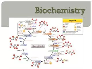
pyruvate kinase phosphoenolpyruvate pyruvate ADP ATP Examples of substrate-level phosphorylations In glycolysis 3-phosphoglycerate kinase 1,3-bisphosphoglycerate3-phosphoglycerate ADP ATP In the citrate cycle thiokinase succinyl coenzyme A succinate + CoA GTP GDP + Pi In skeletal muscle phosphocreatine serves as a reservoir of high-potential phosphoryl groups that can be readily transferred to ATP: creatine kinase phosphocreatine creatine ADP + H+ ATP
ATP in cells • Life expectancy of an ATP molecule is about 2 min. • It must be permanently synthesized • Momentary content of ATP in a human body is about 100 g, but 60-70 kg is produced daily • Adenylate kinase maintains the equilibrium between ATP, ADP a AMP ATP + AMP 2 ADP
Energy status of a cell [ATP]/[ADP] ratio(in most cells 5-200) Energy charge of the cell: The energy charge of most cells ranges from 0.80 to 0.95
Control of metabolism • Metabolism is regulated by controlling • catalytic activity of enzymes allosteric and cooperative effects, reversible covalent modification, substrate concentration • the amount of enzymes synthesis of adaptable enzymes • the accessibility of substrates compartmentalization segregates biosynthetic and degradative pathways, the flux of substrates depends oncontrolled transfer from one compartment of a cell to another • the energy status of the cell of which the energy charge or the phosphorylation potential are used as indexes • communication between cells • hormones, neurotransmitters, and other extracellular molecular signals often regulate the reversible modification of key enzymes
Metabolism of sacharides 1 Celular metabolism of glucose
Transport of Glucose into the cells Molecules of glucose are strongly polar, they cannot diffuse freely across the hydrophobic lipid bilayer Glucose transporters Transmembrane proteins facilitating a transport of glucose 2 main types: GLUT (1-14)* and SGLT** * glucose transporter ** sodium-coupled glucose transporter
GLUT 1-GLUT 14, family of transporters with common structural features (isoforms) but a tissue specific pattern of expresion: 500 AA, 12 transmembrane helices Mechanism of transport: facilitated diffusion (follows concentration gradient, do not require energy)
Differences between the GLUT transporters • affinity to glucose • different way of regulation • tissue specific occurence
Glucose transporters Typ characteristics GLUT 1 Basal glucose uptake (ercs, muscle cells at resting conditions, brain vessels ..) , b GLUT 2 kidney Liver, cells of pancreas GLUT 3 Neurons, placental cells GLUT 4 Muscle, adipocytes – dependent on insulin - GLUT 5 Transport of fructose, small intestine - GLUT 7 Intracelular transport liver
Transport of glucose by GLUT glucose Extracellular space cytosol Two conformational states of transporters
Extracellular space cytosol
GLUT 1 deficiency Inherited deficiency of glucose transporter Transport across the blood brain barrier is reduced The glucose concentration in cerebrospinal fluid is decreased. Moreover GLUT1 is essential also for transport of glucose into neurons and glial cells As glucose is the principal source of fuel to the brain, GLUT1 deficiency causes impaired provision of energy epileptic encephalopathy Observation: decreased level of glucose in liquor Treatment: The only known treatment to date is a very restrictive diet called the ketogenic diet
GLUT 4 carriers are regulated by insulin (muscle, adipocytes) insulin insulin receptor "Sleeping“ GLUT4 in the membranes of endosomes
Binding of insulin to its receptor insulin insulin receptor vesicles move to the membrane
Transport of glucose into the cell insulin receptor Vesicles fuse with the plasma membrane
GLUT 4 receptors– conclusion: The presence of insulinleads to a rapid increase in the number of GLUT4 transporters in the plasma membrane. Hence, insulin promotes the uptake of glucose by muscle and adipose tissue.
Transport of glucose into the epithelial cells of the small intestine and renal tubules cells • SGLT - transporters • Mechanism: cotransport with Na+ • secondary active transport • glucose and Na+ bind to two specific sites of the carrier • they are transported into the cell at the same time (without energy requirement) • Na+ isconsequentlytransported outside the cell by ATPase (active transport - consumption of ATP) • glucose is consequently transported outside the cell by GLUT2
Cotransport of glucose with Na+ Lumen of the small intestine enterocyte
Transporter changes conformation after the binding glucose and Na+ lumen enterocyte
Na+ and glucose enter the cell (symport) lumen enterocyte
Na+/K+-ATPase is located on the capillary side of the cell and pumps sodium outside the cell (active transport) Extracelular space enterocyte
glucose is exported to the bloodstream via uniport system GLUT-2 (pasive transport) glucose enterocyte extracellular space
SGLT1 deficiency Hereditary disturbance in the transport of glucose and galactose Rare disorder (autosomal recessive patern). This failure of active transport prevents the glucose and galactose from being absorbed. Symptoms become apparent in the first weeks of a baby's life. Severe diarrhea leading to life-threatening dehydration, destabilization of the acidity of the blood and tissues (acidosis), stomach cramps. Why diarrhea? The water that normally would have been transported across the brush border with the sugar instead remains in the intestinal tract to be expelled with the stool, resulting in dehydration of the body's tissues and severe diarrhea. 41
Glucose-6-phosphate is formed immediately after the glucose enters a cell: Glucose metabolism in cells glucose + ATP glucose-6-P + ADP Enzymes hexokinase or glucokinase
Glucose phosphorylation glucose The reverse reaction is catalyzed by glucose-6-P phosphatase This occurs only in liver (and in less extent in kidney). glucose ATP Hexokinase, (glucokinase) phosphorylation ADP glucose-6-P
Consequence: • Phosphorylation reaction traps glucose in the cell. • Glc-6-P cannot diffuse through the membrane, because of its negative charges. • Formation of Glc-6-Pmaintains the glucose concentration gradient and accelerates the entry of glucose into the cell. • Only liver (kidney) can convert Glc-6-P back to glucose and release it to blood • The addition of the phosphoryl group begins to destabilize glucose,thus facilitating its further metabolism
Glucose concentration and rate of phosphorylation reaction Concentration of fasting blood glucose Vmax of glucokinase Enzyme activity glucokinase Vmax of hexokinase hexokinase Glucose concentration, mmol/l 10 5 KM - hexokinase KM - glucokinase
glucokinase functions only when the intracellular concentration of glucose in hepatocyte is elevated (after a carbohydrate rich meal) • hexokinase in liver functions at lower concentrations of glucose • Hexokinase is inhibited by glucose 6-phosphate, the reaction product. High concentration of this molecule signals that the cell no longer requires glucose for energy, for storage in the form of glycogen, or as a source of biosynthetic precursors, and the glucose will be left in the blood.
Role of glucokinase in pancreas Glucokinase in - cells of pancreas functions as a blood glucose sensor When blood glucose level is high (after the saccharide rich meal), glucose enters the -pancreatic cells (by GLUT2) and is phosphorylated by glucokinase Increase of energy status in the cell enhances the release of insulin
Glycolysis • Significance: energy gain, formation of intermediates for other processes, includes also the metabolism of galactose and fructose • Occurs in most of tissues • Location: cytoplasma • Reversible, enzyme catalyzed reactions • Three reactions are irreversible Anaerobic glycolysis When oxygen is lacking, pyruvate is converted to lactate Aerobic glycolysis At adequate supply of oxygen, pyruvate is converted to acetylCoA