Download

1 / 59
940 likes | 3.05k Views
OTOLOGIC MANIFESTATIONS OF SYSTEMIC DISEASEs. Isfahan university of medical sciences M.Rogha M.D. 1- Granulomatous & infectious dis. 2- Neoplastic dis. 3- Disease of bone 4- Storage dis. 5- Collagen vascular dis. 6- Immunodeficiency disorder (primary, acquired ). classification.

E N D
OTOLOGIC MANIFESTATIONS OF SYSTEMIC DISEASEs Isfahan university of medical sciences M.Rogha M.D.
1- Granulomatous & infectious dis.2- Neoplastic dis.3- Disease of bone4- Storage dis.5- Collagen vascular dis.6- Immunodeficiency disorder (primary, acquired ) classification
Systemic disease that can affect the ear • 4- Storage and metabolic disease • a. Mucopolysaccharidoses • b. Gout • c. Ochronosis • 5- Collagen vascular and autoimmune disease • 6- Immunodeficiency disorders • a. Primary or congenital • i. Humoral immunodeficiency disorders • ii. Cellular immunodeficiency disorders • iii. Disorders of phagocyte function • iv. Complement system defects • b. Acquired • i. Acquired immunodeficiency syndrome • 1- Granulomatous and infectious disease • a. Langerhans cell histiocytosis (LCH) • b. Tuberculosis • c. Wegners granulomatosis • d. Sarcoidosis • e. Syphilis • f. Lyme disease • g. Mycotic disease • 2- Neoplastic disease • a. Multiple myeloma • b. Leukemia • c. Metastatic neoplasm • d. Paraganglioma • 3- Disease of bone • a. Pagets disease • b. Osteogenesis imperfecta • c. Fibrous dysplesia • d. Osteopetroses • e. Osteitis fibrosa cystica
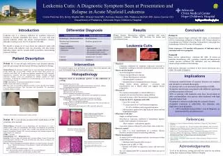
Langerhans cell histiocytosis (LCH)Histiositosis X • - Proliferation of cytologically benign histiocytes • - Etiology and pathogenesis remain unknown • a- unifocaleosinophilic granuloma • - M>F, No systemic manifestation, prognosis is excellent • - Dx: Local curettage + low dose irradiation, Follow-up with radiographic skeletal survey • b- Hand-Schuller-Christian disease • - <5 y, Multifocal osteolytic lesions, With limited Involvement of skin, lymph nodes & viscera • - Systemic manifestations include fever, anorexia, recurrent URTI, anterior cervical lymphadenopathy, otitis media, and hepatosplenomegaly • - Dx: Low dose chemotherapy • C- Letterer-Siwe disease • - <3y, diffuse involvement of multiple organs, manifestations include fever, Seborrheic or eczema-like rash. Oral lesions, lymphadenopathy, hepatosplenomegaly, Multiple bony lesions, diffuse replacement of marrow, and pulmonary infiltration • - Virulent, poor prognosis and high mortality ate • - Dx: Corticosteroids and cytotoxic drugs
Otologic manifestation - Mastoid is a common site of involvement - Otic capsule & facial nerve are relatively resistant - Otorrhea is the most common symotom followed by postauricular swelling, HL, & vertigo - The most common sign is granulation tissue or aural polyps Perforation of the TM, otitis media, otitis external, a fistula between the mastoid and the external canal - Diagnosis of LCH is suggested by an inflammatory disorder of middle ear and mastoid that does not respond to routine antibiotic therapy, bilateral destructive ear disease, an elevated ESR in the absence of acute infection, exuberant granulation tissue after mastoid surgery, and associated skin and systemic lesions - Radiographs show destructive lesions in the mastoid - The diagnosis is established by biopsy - A definitive diagnosis of LCH is made by immunostaining and electron microscopic studies
two lytic lesions of the skull showing beveled edges (arrows) and nonsclerotic margins, which are typical of histiocytosis X.
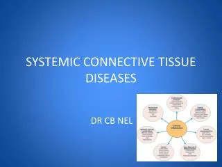
Tuberculosis • Tuberculosis otitis media 0.05% to 0.9% of all cases of COM • Hematogenous or lymphatic or by extension through eustachian tube • TM becomes thickened, CHL due SOME • No pain or tenderness, lymphanopathy in high jugular chain • Multiple small perforation of the TM • The middle ear mucosa appears to be hyperemic with polypoid granulation • Destruction of mastoid tip may result in Bezolds abscess • Definitive diagnosis is made by histopathologic examination Of tissue from the ME or mastoid showing a granulomatous Process with multinucleated giant cells (langerhans cells) and histologic demonstration of acid-fast organisms D.D with wegener granulomatosis and distinguished on the basis of skin test, cultures of the ME, ANCA Dx: Systemic use of standard anti-TB chemotherapy however mastoid surgery may be required to remove sequestrated bone
The TM is intact, but greatly thickened by tuberculous granulation tissue containing the typical epithelioid cells, round cells, and multinucleated giant cells
Wegener’s granulomatosis • A granulomatouse inflammatory process with necrotizing vasculitis • Primarily affects the upper and lower respiratory tract and kidneys but can involve any organ • M=F, mean age 40 y • Common presenting symptom: headache, sinusitis, rhinorrhea, otitis media, fever, arthralgia • Upper airway and sinus involvement in 75% to 90%, pulmonary manifestations(couph, pleuritic chest pain, hemoptysis and nodular or cavitary infiltrates) in 65% to 85% • Glomerulonephritis in 60%-75%, eye involvement (conjunctivitis, iritis, scleritis, proptosis) in 15%-50%, dermatologic findings (necrotic ulceration, vesicles or petechiae) • Laboratory findings: normochromic, normocytic anemia, thrombosytosis, positive RF, hyperglobulinemia particularly IgA, elevated ESR • Positive ANCA test, especially proteinase 3, specificity>95%, sensitivity>90% in active and systemic, in limited or inactive 65% to70%
Continue • The diagnose of WG is made histologically by the presence of necrosis granulomatous inflammation with multinucleated giant cells, vasculitis and microabscess formation • Etiology and pathogenesis unknown, but currently considered to be an autoimmune disease that is perhaps the result of stimulation by an infectious agent (or agents) • Prognosis of WG has dramatically improved from mortality rate of 80% to the current remission rate of >75% • Dx. High doses of corticosteroids, cyclophosphamide or methotrexate for 3 to 6 m followed by maintenance of remission using lower doses of corticosteroids and less toxic immunosuppresants such as azathioprime, methotrexate, trimethoprime-sulfamethoxazole • Otologic manifestation: Middle ear and mastoid are the most common sites within the temporal bone, SOM due to obstruction of the eustachian tube, purulant OM, granulomatous involvement of the ME & mastoid, facial nerve involvement and inner ear can be involved
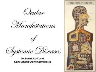
Segmental vasculitis in Wegener granulomatosis with inflammation involving a portion of the arterial wall
SARCOIDOSIS • Chronic multisystem disease of unknown etiology that’s characterized by noncaseating granulomas • It most frequently affects lungs, F>M, and is 10 times more common in blacks, third to fourth decade • Common presenting symptoms: Bilateral hilar adenopathy, cough, granulomatous skin rash • Others: iridocyclitis, keratocojectivitis, peripheral lymphadenopathy, hepatosplenomegaly, cardiac failure, myalgia, and arthralgia • The facial & optic nerves are the most commonly affected cranial nerves • Laboratory findings: Hilar adenopathy in CXR, hypercalcemia, and elevated serum ACE • It has been suggested that the etiology is linked to genetically determined enhancement of the T-helper immune response to a limited number of microbial pathogens
Continue • Spontaneous resolution occurs in many patients • Corticosteroids are beneficial for those with progressive symptoms or with ocular, cardiac or CNS involvement • Infliximab (inhibits release of TNF) has been reported as being effective for some cases that are refractory to other treatments • Otologic manifestation: SNHL, vestibular dysfunction and facial nerve paralysis, or occasionally granulomatous disease of the external or middle ear and mastoid • The facial nerve is the most commonly affected cranial nerve, it is often bilateral, it may resolve spontaneously and is usually involved as part of the triad of uveoparotid fever (Heerfordts syndrome) parotitis, uveitis, facial nerve paralysis, and mild pyrexia
sarcoidosis Bilateral hilar adenopathy and linear parenchymal densities in pulmonary sarcoidosis.
Both congenital and acquired syphilis may affect the middle ear in the late latent and tertiary forms • In late latent form the ME & mastoid affected by rarefying osteitis with leukocytic infiltration • In tertiary the gumma demonstrates obliterative arteritis and central necrosis, A gumma of the ear canal or ME may result in perforation of TM and a granulomatous appearance of mucosa • Definitive diagnosis of syphilis requires a positive serologic test and a histologic demonstration of Treponema pallidum • Syphilis may mimic TB • Heneberts sign (induction of ocular deviation with positive or negative pressure in the external canal) probably due to fibrous adhesion between the stapes footplate and the membranous labyrinth • Dx: Combined antibiotic and corticosteroid SYPHILIS
active round cell osteitis (O)
LYME DISEASE • Multisystem inflammatory disorder that affects skin, nervous system, heart, joints • Spirochete Borreliaburgdorferi • Transmitted by Ixodes Ticks • Primary reservoirs are white-footed mice and white-tailed deer • Three clinical stages are recognized : a- The first stage (early, localized infection) begins 3 to 33 days after a tick bite (erythema migrans), this lesion occurs in 60%-80% of patients and may accompanied by minor constitutional symptoms b- The second stage (early, disseminated infection) occurs within days or weeks after inoculation, symptoms include fever, migratory arthralgia, myalgia, headache, meningismus, generalized lymphadenopathy, malaise, fatigue, and secondary annular skin lesion c-The third stage occurs more than a year after onset and can result in chronic, prolonged arthritis, chronic encephalomyelitis, chronic axonal peripheral polyradiculopathy, keratitis, acrodermatitischronic`atrophicans, localized scleroderma-like lesions
Continue • Inflammatory innate immune responses are critical in the pathogenesis • Diagnosis is based on the recognition of the characteristic clinical features, a history of exposure and detection of a specific antibody to B. burgdorferi • Dx: The spirochete is highly sensitive to doxycycline, other effective antibiotics include amoxicillin, erythromycin, cefuroxime, ceftriaxone, imipenem. Steroids for carditis and arthritis. • Vaccine is now available • Otologic manifestation: Facial nerve paralysis is the most common Otologic manifestation (3% to 11%), bilateral in (25%), in second stage, in all ages and both sexes, acute in onset, return is spontaneous and complete, antibiotics or steroids do not appear to influence the duration or outcome • Lymphocytoma a red and violet nodules occur on the earlobe during the second stage • SNHL, sudden hearing loss, vertigo, meniere-like symptom have been described
Systemic invasive clinical disease reflects some defect in host defense, such as DKA, chemotherapy, AIDS Diagnose is made by biopsy and culture Treatment consists of control of the underlying predisposing condition, surgical debridement of necrotic tissue and Amphotercin- B Otologic manifestation: destruction of the middle ear cleft ensues, often with extention to the surrounding structures , including thrombosis or rupture of the internal carotid artery Other routes: hematogenous embolic dissemination MYCOTIC DISEASE
NEOPLASTIC DISEASE • Multiple myeloma: malignancy of plasmacells derived from B lymphocytes, M>F, 60 y • Severe bone pain, pathologic fractures, renal failure, failure of the bone marrow, hypercalcemia and recurrent infections. • Laboratory findings: M component on serum or urine electrophoresis normochromic, normocytic anemia, hypercalcemia and elevated BUN. • Otologic manifestation: Lytic lesions of the temporal bone and otic capsule, • Symptoms are usually overshadowed by manifestation of diffuse disease • Tx: Autologous stemcell transplantation, thalidomide, bisphosphonate and erythropoietin • Extramedullaryplasmacytoma (soft tissue) and solitary bone plasmacytoma (bone): in younger individual, M component in 30%, indolent course, survival rates of 10y or more • Dx: local radiotherapy (4000 cGY), • Periodic evaluation should be performed to detect conversion to multiple myeloma
large lytic destructive lesion (arrows) of the clivus (CL), petrous temporal bone, middle ear, and jugular foramen area caused by multiple myeloma
Coronal CT scan with contrast enhancement and a soft tissue technique shows a slightly enhancing mass that has destroyed the mastoid bone and extends to the posterior fossa (PF) and upper neck (UN).
Common in thesubmucosaof the pneumatized areas of middle ear and mastoid and bone marrow of the petrous apex • Secondary bacterial infection due to immunocompromised state or chemotherapy, hemorrage in ME, mastoid or inner ear • Clinical manifestation: ME effusion, acute and chronic suppuration in the ME and mastoid, thickening of the TM, CHL, SNHL, vertigo, facial paralysis, skin lesions in the external auditory canal • Granolocytic sarcoma or chloroma: exteramedullarytumore in AML or CML • Management is by local irradiation and chemotherapy LEUKEMIA
PARAGANGLIOMA • Is the most common neoplasm after the acoustic neuroma • Divided into two groups: the glomus tympanicum and glomus jugulare • Symptom: • The glomus tympanicum appears with pulsatile tinnitus and a CHL • The glomus jugulare appears late, after considerable growth and bony destruction, may cause a neurologic defect in CNs IX to XII, facial nerve paresis caused by tumor extension into the mastoid, or SNHL caused by bony erosion of the labyrrinth • Both may erode the TM and presenting by bleeding mass • 10% of nonfamilial and 50% of familial have at least one additional Lesion • A few PG, both benign and malignant may secrete catecholamines • A history of headache, hypertension and flushing • Dx: surgery, radiotherapy is useful for management of recurrences and unresectable lesions
METASTATIC NEOPLASM • Hematogenous dissemination • The most common sites: • Breast, lung, prostate, skin • Petrous apex and internal auditory canal • The otic capsule relatively resistant • CHL, pain, SNHL, vertigo, facial paralysis • In meningial carcinomatosis unilateral or bilateral SNHL is a common presenting symptom, diagnosis is made by cytology of the CSF
metastatic breast adenocarcinoma showing a large lytic lesion (arrows) destroying the mastoid.
PAGETS DISEASE (osteitis deformans) • Osteolytic and osteoblastic changes affect the axial skeleton • AD, 3% of the population, 40y old and older, M>F • Enlarging skull, progressive kyphosis, deformities of the pelvis, femur, tibia • Etiology is uncertain, slow virus infection have suggested • Tx: bisphosphonate, calcitonin, mithramycin, ipriflavone, gallium nitrate • Otologic manifestation: HL, tinnitus, mild vestibular dysfunction, facial nerve is spared • HL 5% to 44%, SN, mixed or rarely conductive • D.D: otosclerosis, paget is late in onset, old age, greater SNHL, enlarged calvaria, enlargement of the superficial temporal artery, elevated serum ALP
Paget's disease. There is diffuse expansion of the skull table and involvement of both temporal bones, with patchy demineralization.
Lateral skull radiograph in a patient with Paget's disease. Findings include thickening of the skull table, multiple patchy densities, and platybasia.
The pagetic bone encroaches on the posterior margin (arrow) of the internal auditory canal (IAC). The mastoid is largely replaced by pagetic bone
OSTEOGENESIS IMPERFECTA • Type I through IV • Type I: AD, mildest form, blue sclera, nondeformingfructures, normal stature, HL in 30%-50% • Type II: most severe, multiple fracture in uterus, stillbirth, AR or sporadic • Type III: multiple fracture, bone deformity, HL in 50% • Type IV: AD, similar to type I except that the sclera are white, HL in 10%-30% • Tx: management of fractures, orthopedic surgery, bisphosphonate • Otologic manifestation: SNHL in 40%, high correlation with gray or white sclera, CHL accompanies blue sclera, CHL reflects structural change in the ossicles, microfractures of the manuberium, fragility of the long process of the incus, fracture or resorption of the crura of the stapes • Rehabilitation by amplification or surgery • Stapedectomy can give similar results to otosclerosis
FIBROUS DYSPLASIA • Benign, chronic, slowly progressive, unknown etiology • Replacement of normal bone with fibrous tissue and woven bone • 7% as part of Albrights syndrome (bony lesions, abnormal pigmentation, endocrine dysfunction, precocious puberty in women) • 70% monostotic form: most common, skull, ribs, femur, tibia, may become quiescent at puberty • 23% polystotic form: skull lesions in more than 50%, can continue to progress • Clinical manifestation: bony deformity, pathologic fracture, cranial nerve palsy • Normal serum calcitonin and phosphorus levels, elevated serum ALP in polystotic form • Radiographic finding: radiolucent area, ground-glass appearance • Otologic manifestation: progressive narrowing of the external auditory canal with CHL is the most common (80%), facial nerve paralysis, SNHL, vertigo • Management is symptomatic, radiotherapy is contraindicated
Lateral radiograph of the skull of a patient with fibrous dysplasia showing lytic (L) and fibrous (F) phases of disease. Spicules of new bone are responsible for the ground-glass appearance of the fibrous phase.
Coronal tomographic radiograph of a patient with fibrous dysplasia. New bone formation causes a dense appearance of the involved left temporal bone.
OSTEOPETROSES • Rare genetic disorder, greatly increased bone density, • Defective function of osteoclasts, • Malignant osteopetrosis ; AR, high mortality rate, anemia, thrombocytopenia, hepatosplenomegaly, susceptibility to infection, encroachment of the neural foramina, optic atrophy, facial paralysis, SNHL, hydrocephalus, MR, and death • Otologic manifestation: mastoid is non pneumatized, inner ear normal, herniation of facial nerve a consistent finding, recurrent episodes of AOM, SOM, CHL, SNHL, unilateral or bilateral facial nerve paralysis • Tx: symptomatic, decompression of the facial nerve
OSTEITIS FIBROSA CYSTICA • Von Recklinghausens disease • Excess parathormone • Osteoclastic bone resorption, marrow fibrosis, bone cysts, bone pain, and fractures • In most cases is caused by hyperparathyroidism due to an adenoma • Involvement of temporal bone is very rare, the otic capsule is replaced by abnormal bone, SNHL has been attributed to osteitisfibrosa
MUCOPOLYSACCHARIDOSES • MPS an inherited deficiency of one of several lysosomal enzymes that degrade MPS • Classified into seven types, all are AR except for Hunters syn (MPS II) which is X-linked recessive • Management is supportive and symptomatic • MPS III: Hurlers syn, accumulation of heparan sulfate, corneal clouding, abnormal facies, hepatosplenomegaly, MR, joint stiffness, and hernias • MPS II: Hunters syn, accumulation of heparan sulfate and dermatan sulfate, similar to hurler, but corneal clouding is not seen • MPS IV: Moquios syn, spondyloepiphyseal dysplasia • Otologic manifestations: CHL( Eustachian tube dysfunction & thickening of the mucosa ), SNHL ( may be a result of abnormal metabolism of the inner ear).
GOUT • Deposition of crystals of monosodium urate within joint space & cutaneous structures • Serum urate level>7mg/dl, risk factors include: alcohol use, exposure to loop diuretics, hypertension & renal insufficiency • Clinical manifestation: acute gouty arthritis, tophi, urate urolithiasis, and gouty nephropathy • Otologic manifestation: Tophaceous deposits in the helical rim of the pinna, asymptomatic • Treatment: bed rest, analgesics, colchicine, probencid, allopurinol
Ochronosis • Ochronosis is a rare disease that is caused by an inherited lack of the enzyme homogentisic acid oxidase. • The presence of homogentisic acid in urine is called alkaptonuria. The result of this inborn error of metabolism is the deposition of a dark pigment in tissues that are rich in collagen. • Patients often present with symptoms and signs during the third decade of life. • Manifestations include ochronoticarthropathy, ocular and cutaneous pigmentation, obstruction of the genitourinary tract by ochronotic calculi, and cardiovascular manifestations as a result of ochronosis affecting the aortic valve. • Ochronosis has manifestations in the external ear; cartilage is a site of predilection for the deposition of the pigment of ochronosis. Blue or mottled-brown macules can appear on the pinna and in other areas of the head and neck, including the nose, buccal mucosa, tonsils, pharynx, larynx, and esophagus.