Download
1 / 51
1.64k likes | 5.52k Views
MOLECULAR BASIS OF CANCER. Dr Kiran H S ASSISTANT PROFESSOR. OVERVIEW. Fundamental Principles Essential alterations for malignant transformation Conclusion References. Carkinos - CRAB. CANCER – Uncontrolled growth of cells. history. Hippocrates – Tumor Resembled a Crab

E N D
MOLECULAR BASIS OF CANCER Dr Kiran H S ASSISTANT PROFESSOR
OVERVIEW • Fundamental Principles • Essential alterations for malignant transformation • Conclusion • References
Carkinos - CRAB CANCER – Uncontrolled growth of cells
history • Hippocrates – Tumor Resembled a Crab • Oldest documented - 1500 BC in Egypt 8 cases of tumors in the Breast.
Research identifying carcinogens, radiation therapy , chemotherapy and better means of diagnosis were discovered. • Today, we are able to cure some types of cancer, and research is ongoing.
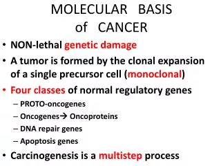
Molecular basis of cancer Fundamental principles: 1.Nonlethal genetic damage: chemicals • Acquired radiation viruses • Inherited in the germline
2. Tumors are monoclonal • A tumor is formed by the clonal expansion of a single precursor cell that has incurred the genetic damage. • Assessed in women who are heterozygous for polymorphic X – linked markers , such as androgen receptors.
3. Principle targets of genetic damage: • Proto – oncogenes : Growth promoting (mutant alleles are dominant) • Tumor suppressor genes –Growth inhibiting (both normal alleles suppressed) Loss of gene function caused by damage to a single allele - Haploinsufficiency.
Genes that regulate apoptosis • Genes involved in DNA repair
4.Carcinogenesis is a multistep process at both phenotypic and genetic levels resulting from accumulation of multiple mutations. • Tumor progression – Acquire malignant potential • Molecular level - accumulation of genetic lesions. • Most malignant tumors are monoclonal in origin , constituent cells are heterogenous by the time they become malignant.
Essential Alterations for malignant transformation • Self-sufficiency in growth signals • Insensitivity to growth-inhibitory signals • Evasion of apoptosis • Limitless replicative potential • Sustained angiogenesis • Ability to invade and metastasize • Defects in DNA repair
Nature of genes in biologic alteration Proto – Oncogenes: • Code for cellular proteins which regulate normal cell growth and differentiation. • Proteins encoded by proto-oncogenes participate in control of cell growth: 1. Growth factors 2. Receptors for growth factors 3. Signal transducers 4. Transcription factors 5. Cell cycle receptors
Mutations convert proto – oncogenes to oncogenes involved in tumor development. • How do these normally “civilized” growth factors turn into “enemies” within ? • Point mutation • Amplification • Overexpression • Gene rearrangements
Growth factors • Proliferation of normal cells. • Paracrine action : Growth factors made by one cell type act on neighboring cell - Proliferation • Autocrine Loop – Ability of cancer cells to synthesize same growth factors to which they are responsive.
RET protein : • Receptor for glial cell line derived neurotrophic factor. • Normally expressed – Parafollicular C cells of thyroid, adrenal medulla, parathyroid cell precursors. Point mutation – MEN 2A & 2B,Familial MCT • RET Somatic rearrangements – Sporadic MCT
MEN 2A – Point mutation in extracellular domain. MCT , Adrenal & Parathyroid tumors. • MEN 2B – Point mutation in cytoplasmic domain. Thyroid & adrenal tumors without parathyroid. Also – Ganglioneuroma , Marfanoid
EGF receptor family : • ERBB1 Over expression – 80% SCC lung 50% glioblastomas 80 % – 100 % head & neck tumors. • ERBB2 (HER-2/NEU) Amplification – 25% breast cancers AdenoCa-ovary,lung,salivary gland • Monoclonal antibodies specific to ERBB2 - targeted therapy.
RAS Oncogene : • Discovered in transforming retroviruses • 3 in human genome - HRAS , KRAS , NRAS • Point mutations in RAS family genes – most common abnormality of proto – oncogenes in humans. • Frequency – varies with different tumors.
90% cases – Pancreatic AdenoCa & CholangiCa • 50% - Ca colon , endometrium , thyroid • 30% - Adenocarcinoma lung , Myeloid leukemias • KRAS – Colon , Lung , Pancreas HRAS – Bladder and Kidney NRAS – Melanoma , Leukemias
Tumor suppressor genes • Oncogenes : Drive the proliferation of cells by gaining function. • Tumor suppressor genes : Loss of function leads to cell proliferation . Recognize genotoxic stress and respond by shutting down proliferation. Cause cell to enter postmitotic differentiated pool without replicative potential.
Protein products : Transcription factors Cell cycle inhibitors Signal transduction molecules Cell surface receptors Regulators of cellular responses to DNA damage
RB gene: • First & prototypic tumor suppressor gene. • Discovered by studying a rare disease – Retinoblastoma. 60% sporadic • Retinoblastoma 40% familial
Knudson’s double hit hypothesis • Two mutations involving both alleles of RB at chr locus 13q14 are required to produce retinoblastoma. • Cancer develops when the cell becomes homozygous for the mutant allele / when cell loses heterozygosity for the normal RB gene (LOH)
RB protein : • Product of RB gene. • Key role in regulating cell cycle. • Plays impt role in deciding if the cells should enter or exit the cycle at G1 phase,differentiate or die.
p53 : Guardian of the genome: • Chr 17p13.1 • Molecular Policeman – prevents propagation of genetically damaged cells. • MC target for genetic alteration in human tumors. • > 50% of human tumors – have mutation in this gene. • It is a transcription factor – can sense cellular stress ( DNA damage, shortened telomeres,hypoxia )
p53 prevents neoplastic transformation : • Activation of temporary cell cycle arrest ( quiescence ) • Induction of permanent cell cycle arrest (senescence ) • Triggering of programmed cell death (Apoptosis )
NF1 : • Individuals inherit one mutant allele of NF1 gene. ( located on 17q11.2) • Numerous benign neurofibromas,optic nerve gliomas,Lischnodules,café au lait spots • Neurofibromin : protein product of NF1 Regulate signal transduction through RAS protein .( converts active RAS to inactive)
NF 2: • Mutation in NF 2 – B/l Acoustic nerve shwannomas , meningiomas , ependymomas. • Less common than NF 1 • Gene located on chr 22q12. • Gene product – Merlin - Cells lacking this are insensitive to normal growth arrest signals.
WT1: • Located on chr 11p13 • Transcriptional activator of genes involved in renal & gonadal differentiation. • Regulates mesenchymal – epithelial transition in kidney. • Mutation WT1 – Wilm’s tumor • WT2 , chr 11p15 : Beckwith – Wiedmann syndrome ( macroglossia,hemihypertrophy, omphalocele)
Evasion of apoptosis • Mutations in genes regulating apoptosis also play a critical role in neoplasia. • Various signals trigger apoptosis • Large family of genes which regulate apoptosis have been identified. • Two pathways activate apoptosis : Intrinsic and extrinsic.
Telomerase and cancer • Loss of ability of cell to divide and become senescent is mainly due to shortening of telomere. • Telomeres – Short repeated sequences of DNA at the ends of chromosomes. • Telomere length – maintained by Telomerase. Highest activity in germ cells Less in stem cells Undetectable in somatic tissues.
Cancers : Telomere length is maintained Upregulation of telomerase Telomere lengthening ( 85% - 90% of cancers) • Eg : Colonic adenoma – Adenocarcinoma Early lesions – low telomerase activity. Malignant lesions – high telomerase activity.
angiogenesis • Tumors require vascularisation for their growth. • Cancer cells stimulate neoangiogenesis. • Tumor vasculature is abnormal – leaky vessels with haphazard pattern of arrangement. Supply of nutrients • Dual effect Release GFs- growth of adjacent tumor cells.
Balance b/w angiogenesis promoters (bFGF,VEGF) and inhibitors (Angiostatin,endostatin,vasculostatin , thrombospondin) • Factors produced by – tumor cells, macrophages, stromal cells associated with tumor. • Angiogenic switch - Controlled by physiologic factors. • Pro and anti-angiogenic factors – regulated by genes mutated in cancer.(p53)
Invasion and metastasis • Biologic hallmark of malignant tumors • Major cause of cancer related morbidity & mortality. • Metastatic Cascade Invasion of ECM Vascular dissemination,homing of tumor cells,colonization.
Invasion of ECM: • Initiates metastatic cascade • Several steps Loosening of intercellular junctions Degradation of ECM Attachment to novel ECM components Migration of tumor cells.
Loosening of cell interactions: • Alteration in itercellular adhesion molecules. • E- cadherin Down-regulation: Reduces cell adhesion Seen in majority of epithelial tumors eg: Adenocarcinoma of colon , breast
Degradation: • Basement membrane and interstitial connective tissue. • Tumor cells elaborate proteases • Belong to different families : MMPs, Cathepsin D, Urokinase plasminogen activator.
Matrix metalloproteinases: • Recognised as important components of tumourigenesis. • Play role in turnover of cellular receptors, growth factors, cytokines, chemokines and ECM. • Produced by tumor cells and stromal cells. • In humans there are 23 MMPs • Eg: MMP9 – stimulates release of VEGF
Vascular Dissemination: • Tumor cells tend to aggregate. (homotypic /heterotypic) • Bind to coagulation factors. • Extravasation at distant site – adhesion molecules eg: CD44 expressed on T lymphocyte
Organ tropism: • Tumor cells have adhesion molecules whose ligands are expressed preferentially on endothelial cells of target organs. • Chemokines – decide target tissue eg: Ca breast – CXCR4 & CCR7 • Target tissue – unfavourable for tumor growth. Eg : Skeletal muscle.
references • StrickerTP,KumarV.Neoplasia.Robins and Cotran Pathologic Basis of Disease 2010:259-329. • Kumar V,FaustoN,AbbasK.Tissue Renewal, Repair,andRegeneration.Robins and Cotran Pathologic Basis of Disease 2010:79-110. • LibermanW,LebovitzR.Neoplasia.Anderson’s pathology tenth edition:500-544. • Murphy G.Matrixmetalloproteinases in neoplastic progression. Recent advances in histopathology 22: 81 – 90. • Buda A,PignatelliM.Micrsatellite instability and neoplasia. Recent advances in histopathology 20.167 – 181.