Download
1 / 12
120 likes | 833 Views
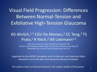
Visual Field Progression: Differences Between Normal-Tension and Exfoliative High-Tension Glaucoma. KG Ahrlich, 1,3 CGV De Moraes, 2 CC Teng, 2 TS Prata, 2 R Ritch, 2 JM Liebmann 1,2. 1 New York University School of Medicine, New York, NY

E N D
Visual Field Progression: Differences Between Normal-Tension and Exfoliative High-Tension Glaucoma KG Ahrlich,1,3 CGV De Moraes,2 CC Teng,2 TS Prata,2 R Ritch,2 JM Liebmann1,2 1New York University School of Medicine, New York, NY 2Einhorn Clinical Research Center, New York Eye & Ear Infirmary, New York, NY 3Manhattan Eye, Ear, and Throat Hospital, New York, NY Supported by the ASCRS Foundation and the Ephraim and Catherine Gildor Research Fund of the New York Glaucoma Research Institute. The authors have no financial interest in the subject matter of this poster.
Introduction • The relative importance of IOP-dependent and IOP-independent risk factors varies among individuals and forms of glaucoma. • Exfoliative glaucoma (XFG) is characteristically associated with elevated IOP (exfoliative high tension glaucoma, XHTG), and IOP-dependent factors are thought to play a central role in disease onset and progression. • Glaucomatous eyes with an IOP in the statistically normal range (normal-tension glaucoma, NTG) are less dependent on IOP for disease onset and progression. • It remains unclear whether the same pattern and rates of glaucomatous visual field deterioration are present in both NTG and XHTG.1-8
Purpose • To compare the pattern, location, and rate of visual field (VF) loss in NTG and XHTG.
Methods • The Glaucoma Progression Study (GAPS) consists of 43,660 consecutive subjects (132,512 VF tests) evaluated in a glaucoma referral practice from January 1999 to December 2008. • Subjects with glaucomatous optic neuropathy, repeatable VF loss, ≥5 SITA-Standard VF examinations, and NTG or HTG, were enrolled. If both eyes were eligible, one was selected randomly. • NTG was defined as glaucomatous VF loss and all known IOP measurements ≤21 mmHg. • HTG was limited to exfoliative glaucoma (XFG), defined as glaucomatous VF loss, untreated IOP >21 mmHg, and the presence of exfoliation material on the pupillary margin and/or on the anterior lens capsule.
Methods • VISUAL FIELD ANALYSIS • Automated pointwise linear regression (PLR) analysis was performed using Progressor™ (Version 3.3, Medisoft, Inc., London, UK), providing slopes (decibels [dB]/year) of progression globally and locally for each point based on threshold maps, as well as significance (p-values). • The number and location of the significantly progressing points was compared with the division of VF sectors described by Garway-Heath et al.9 This information was used to establish the most common location of progressing points in each group.
Methods • CLINICAL DATA • Baseline central VF loss was defined by the presence of at least one point with p<0.01 within the four central-most points of the pattern deviation graph in the two consecutive baseline tests. • Progression was defined as the presence of a test point with a slope of sensitivity over time >1 dB loss/year, with p<0.01. For edge points, a stricter slope criterion of >2 dB loss/year (also with p<0.01) was used. • Paracentral progression was defined as progression of any of the points adjacent to the four central-most points of the VF (i.e., within the 12 central-most points).
Results Table 1. Baseline characteristics of the studied population. VF=visual field, NTG=normal-tension glaucoma, XHTG=exfoliative high-tension glaucoma, CCT=central corneal thickness, IOP=intraocular pressure. *Includes: hypertension, coronary ischemia, stroke.
Results Table 2. Intercurrent characteristics of the studied population. VF=visual field, NTG=normal-tension glaucoma, XHTG=exfoliative high-tension glaucoma, CCT=central corneal thickness, IOP=intraocular pressure. 1Values are adjusted for differences in age, CCT, and mean IOP between groups.
Results Figure. Mapping of the location of significant visual progression in glaucomatous eyes that reached a progression endpoint (modified from Garway-Heath et al.9 Significant progression was defined by any test point with a slope >1.0 dB loss/year with p<0.01 (or >2.0 db loss/year for edge points). A, NTG; B, XHTG.
Discussion • We optimized the evaluation of the velocity and pattern of VF progression associated with IOP by comparing a group of patients with non-IOP-dependent factors (NTG) and one in which IOP is believed to play a predominant role (XHTG). • XHTG and NTG eyes progress at a similar global rate after adjustment for differences in CCT, IOP, and age. However, NTG eyes progress more often in the central field, independent of other factors. • The most important factor associated with paracentral progression among eyes that reached a progression endpoint was the diagnosis of NTG. • The results of our analysis of VF progression correlate well with previous studies of NTG and XHTG, despite our use of trend analysis by PLR.10,11 • Our map (figure) shows that in eyes with statistically elevated IOP, superior and inferior arcuate areas progress faster, whereas the central field may be more influenced by IOP-independent factors. This requires further clarification.
Conclusion • NTG eyes tended to show a faster progression rate in the central field, but rates of global VF loss are similar between treated NTG and XHTG patients. • Greater surveillance of the central field in NTG may be warranted, with more widespread use of alternative methods to follow NTG patients, including: • visual field strategies assessing the central ten degrees • multifocal visual evoked potential techniques • microperimetry
References • Hitchings RA, Anderton SA. A comparative study of visual field defects seen in patients with low-tension glaucoma and chronic simple glaucoma. Br J Ophthalmol. 1983;67:818-821. • Caprioli J, Spaeth GL. Comparison of visual field defects in the low-tension glaucomas with those in the high-tension glaucomas. Am J Ophthalmol. 1984;97:730-737. • Chauhan BC, Drance SM, Douglas GR, Johnson CA. Visual field damage in normal-tension and high-tension glaucoma. Am J Ophthalmol. 1989;108:636-642. • Araie M, Yamagami J, Suziki Y. Visual field defects in normal-tension and high-tension glaucoma. Ophthalmology. 1993;100:1808-1814. • Araie M. Pattern of visual field defects in normal-tension and high-tension glaucoma. CurrOpinOphthalmol. 1995;6:36-45. • Thonginnetra O, Greenstein VC, Chu D, Liebmann JM, Ritch R, Hood DC. Normal versus high tension glaucoma: a comparison of functional and structural deficits. J Glaucoma. 2009 [Epub ahead of print]. • Motolko M, Drance SM, Douglas GR. Visual field defects in low-tension glaucoma. Comparison of defects in low-tension glaucoma and chronic open angle glaucoma. Arch Ophthalmol. 1982;100:1074-1077. • King D, Drance SM, Douglas G, Schulzer M, Wijsman K. Comparison of visual field defects in normal-tension glaucoma and high-tension glaucoma. Am J Ophthalmol. 1986;101:204-207. • Garway-Heath DF, Poinoosawmy D, Fitzke FW, Hitchings RA. Mapping the visual field to the optic disc in normal tension glaucoma eyes. Ophthalmology. 2000;107:1809-1815. • Drance S, Anderson DR, Schulzer M. Risk factors for progression of visual field abnormalities in normal-tension glaucoma. Am J Ophthalmol. 2001;131:699-708. • Puska P. Unilateral exfoliation syndrome: conversion to bilateral exfoliation and to glaucoma: a prospective 10-year follow-up study. J Glaucoma. 2002;11:517-524.