Download
1 / 105
1.06k likes | 1.41k Views
The Muscular System سیستم عضلی. EDO 001.10. 07/05/2012. The Muscular System سیستم عضلی. Learning Objective: The Physical Therapy Technician will gain an understanding of the anatomy and physiology of the muscular system and its purpose within the body .

E N D
The Muscular Systemسیستم عضلی EDO 001.10 07/05/2012
The Muscular Systemسیستم عضلی Learning Objective: The Physical Therapy Technician will gain an understanding of the anatomy and physiology of the muscular system and its purpose within the body. Estimated Time to Complete: 140 minutes. زمان تعین شده برای تکمیل این لکچر: 140 دقیقه AFAMS
Teaching Pointsنکات تدریس • How skeletal muscle produce movement. • How skeletal muscle are named. • Principal skeletal muscles. • Homeostatic imbalances related to the muscular system. • Surface Anatomy. • Questions. • In-class assignment.
The Muscular System Movement سیستم عضلات • گروپهای بزرگ عضلات اسکلیتی • حرکات در مفاصل مخصوص چطور واقع میگردد. • منشآ، ارتکاز و دخول، وظایف، و تعصیب تمام عضلات بزرگ را بیاموزید. • برای تداوم مراقبت های صحی و توانایی فزیکی محصلین مهم میباشد. • Skeletal muscle major groupings • How movements occur at specific joints • Learn the origin, insertion, function and innervation of major muscles • Important to allied health care and physical rehabilitation students
Muscle Attachment Sites: Origin and Insertionمحلات اتصال عضله: منشاء و دخول • Skeletal muscles shorten & pull on the bones they are attached to • Origin is the bone that does not move when muscle shortens (normally proximal) • Insertion is the movable bone (some 2 joint muscles) • Fleshy portion of the muscle in between attachment sites = belly
عضله اسکلیتی به عظام که آنها وصل است باعث کوتا و کش شدن آنها میگردد. • زمانیکه عضله کوتاه میگردد منشاء در حقیقت عظم است که حرکت نمی کند (بشکل نارمل قریبه) • دخول در حقیقت عظم قابل حرکت است (بعضی ها دو مفصل عضلی است) • قسمت فربه عضله در حقیقت بین ناحیه اتصالی میباشد= بطن
Muscles that Move the Headعضلات که باعث حرکت رأس میگردد • Sternocleidomastoid muscle: • arises from sternum & clavicle & inserts onto mastoid process of skull • innervated by cranial nerve XI (spinal accessory) • contraction of both flexes the cervical vertebrae & extends the head • contraction of one laterally flexes the neck and rotates the face in opposite direction
عضله عنق یا سترنو کلیدو ماستوید • این عضله در حقیقت از عظم قص و ترقوه منشاء گرفته و از قسمت بارزه ماستوید داخل جمجمه میگردد. • توسط عصب قحفی یازدهم (نخاعی فرعی) تعصیب شده است. • تقبض این هر دو عضله باعث قبض فقرات رقبی و بسط رأس میگردد. • و تقبض یکی از آنها عنق را بطرف وحشی قبض نموده و وجه را به جانب مخالف تدور میدهد.
Surface Anatomy • A branch of gross anatomy that examines shapes and markings on the surface of the body as they relate to deeper structures. • Essential in locating and identifying anatomic structures prior to studying internal gross anatomy. • Health-care personnel use surface anatomy to help diagnose medical conditions and to treat patients.
Surface Anatomy • four techniques when examining surface anatomy • visual inspection • directly observe the structure and markings of surface features • palpation • feeling with firm pressure or perceiving by the sense of touch) • precisely locate and identify anatomic features under the skin • percussion • tap sharply on specific body sites to detect resonating vibrations • auscultation • listen to sounds emitted from organs
Triangles of the Neck • Neck/cervical region/cervix is a complex region that connects the head to the trunk. • Spinal cord, nerves, trachea, esophagus, and major vessels traverse this highly flexible area. • Neck contains other organs and several important glands. • Neck can be subdivided into anterior, posterior, and lateral regions.
The Anterior Region of the Neck • Has several palpable landmarks, including the larynx, trachea, and sternal notch. • The larynx. • found in the middle of the neck • composed of multiple cartilages • thyroid cartilage • “Adam’s apple” • Inferior to the larynx are the cricoid cartilage and trachea. • Terminates at the sternal (jugular) notch of the manubrium and the left and right clavicles.
The Clavicles • Paired clavicles and the sternal (jugular) notch represent the border between the thorax and the neck. • On the superior anterior surface where they extend between the base of the neck on the right and left sides laterally to the shoulders. • Left and right costal margins of the rib cage form the inferior boundary of the thorax. • Costal angle (costal arch) is where the costal margins join to form an inverted V at the xiphoid process. • On a thin person, many of the ribs can be seen. • Most of the ribs (with the exception of the first one) can be palpated.
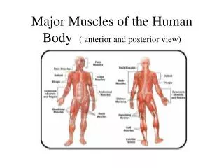
Muscles of Abdominal Wallعضلات جدار بطن • Notice 4 layers of muscle in the abdominal wall
Muscles of Abdominal Wall عضلات جدار بطن • 4 جوره عضلات ورق یا شیت مانند • رکتوس ابدومین= تمایل به عمود دارد. • عضلات مایل داخلی و خارجی و بطنی مستعرض • تدور بدون پیچیده شده تا جدار قدامی بدن را بسازد. • پوش عضله رکتوس و لینا البا را میسازد. • اربطه مغبنی از قسمت قدامی علوی برامده گی حرقفی شروع و به جانب جسم عانه میرود. • کانال مغبنی= یک راه عبوری از حوصله تا به جسم جدار عضلی باز بوده که بشکل یک حلقه مغبنی سطحی دیده میشود. • فتق مغبنی= پاره گی یا تجرید جدار بطن اجازه میدهد تا بخشی از امعأی رقیقه خارج گردد (که بیشتر نزد مردها معمول است.) • 4 pairs of sheet like muscles • rectus abdominis = vertically oriented • external & internal obliques and transverses abdominis • wrap around body to form anterior body wall • form rectus sheath and linea alba • Inguinal ligament from anterior superior iliac spine to upper surface of body of pubis • Inguinal canal = passageway from pelvis through body wall musculature opening seen as superficial inguinal ring • Inguinal hernia = rupture or separation of abdominal wall allowing protrusion of part of the small intestine (more common in males)
Transverse Section of Body Wallبخشی مستعرض جدار بدن • Rectus sheath formed from connective tissue aponeuroses of other abdominal muscles as they insert in the midline connective tissue called the linea alba
پوش ریکتوس از انساج منظم ترکیب یافته و پوش وتری دیگر عضلات بطن به مجرد که داخل خط متوسط انساج منظم میگردد بنام لینا البا یاد میگردد.
Muscles Used in Breathingعضلات که در فعل تنفس استعمال میگردد • در هنگام فعل تنفس ضرور است تا اندازه قفس صدری تغیر نماید. • در زمان شقیق، جوف صدر از نظر اندازه بزرگ میگردد. • عضله خارج بین الضلعی اضلاع را بلند مینماید. • حجاب عاجز تقلص نموده و شکل گنبد مانند آن هموار میگردد. • در زمان زفیر، جوف صدر از نظر اندازه کوچک میگردد. • عضله داخل بین الضلعی در جریان زفیر جبری استعمال میگردد. • حجاب عاجز توسط عصب فرینیک (حجاب حاجزی) (C3-C5) تعصیب شده اما بین اضلاع توسط اعصاب صدری نخاعی (T2-T12) تعصیب شده است. • Breathing requires a change in size of the thorax • During inspiration, thoracic cavity increases in size • external intercostal lift the ribs • diaphragm contracts & dome is flattened • During expiration, thoracic cavity decreases in size • internal intercostal mm used in forced expiration • Diaphragm is innervated by phrenic nerve (C3-C5) but intercostals innervated by thoracic spinal nerves (T2-T12)
Muscles Used in Breathingعضلات که در فعل تنفس استعمال میگردد
SURFACE ANATOMY OF THE LUNGS Where to stick your stethoscope.
Surface anatomy of the lungs • Apices: 2.5cm above the medial 1/3rd of clavicle. • Medial border: pass behind the sternoclavicular joint and down to the sternal angle. On left the border deviates to the left 3cm at 4th costal cartilage, then continues down to the 6th. • The lower borders of the lung are: • T6 - mid-clavicular line (anterior) • T8 - mid-axillary line (Lateral) • T10 - posteriorly
Surface anatomy (cont) • The lower borders of the pleura are: • T8 - mid-clavicular line • T10 - mid-axillary line • T12 - posteriorly • Lung Fissures: • Oblique fissure runs from the spinal process of T3 posteriorly to the level of T6 anteriorly; • The transverse fissure is on the right at T4. • Lobes: Lobes are created by the fissures. • Right lung: upper, middle and lower lobe • Left lung: upper and lower lobe.
Surface anatomy (cont) • The anterior aspect of the chest wall: • Upper lobe – right and left. • T4-T6 is the middle lobe – right only. • The axilla and lateral aspect of the chest wall: • upper, middle (right) and lower lobes. • The posterior aspect of the chest wall: • Lower lobes – right and left.
Surface anatomy of the lungs Front of thorax, showing surface relations of bones, lungs (purple), pleura (blue), and heart (red outline). Side of thorax, showing surface markings for bones, lungs (purple), pleura (blue), and spleen (green).
Thorax • The superior portion of the trunk sandwiched between the neck superiorly and the abdomen inferiorly. • Consists of the chest and the “upper back.” • On the anterior surface of the chest are the two dominating surface features of the thorax. • the clavicles and the sternun
The Sternum • Palpated readily as the midline bony structure in the thorax. • The manubrium, the body, and the xiphoid process may also be palpated. • Sternal angle can be felt as an elevation between the manubrium and the body. • Sternal angle is clinically important because it is at the level of the costal cartilage of the second rib. • it is often used as a landmark for counting the ribs
The Abdomen • On the anterior surface of the abdomen, the umbilicus (navel) is the prominent depression or projection in the midline of the abdominal wall. • In the midline of the abdominal anterior surface is the linea alba, a tendinous structure that extends inferiorly from the xiphoid process to the pubic symphysis. • The left and right rectus abdominis muscles and their tendinous insertions are referred to as “six-pack abs.” • The superior aspect of the ilium (iliac crest) terminates anteriorly at the anterior superior iliac spine. • Attached to the anterior superior iliac spine is the inguinal ligament, which forms the lower boundary of the abdominal wall.
The Inguinal Ligament • Terminates on a little anterior bump on the pubis called the pubic tubercle. • Superior to the medial portion of the inguinal ligament is the superficial inguinal ring. • a superficial opening in the lower anterior abdominal wall • represents a weak spot in the wall • can be palpated to detect an inguinal hernia
Lever Systems and Leverageسیستم اهرمی و شیوه بکار برده اهرمی • Muscle acts on rigid rod (bone)that moves around a fixed point called a fulcrum • Resistance is weight of bodypart & perhaps an object • Effort or load is work doneby muscle contraction • Mechanical advantage • the muscle whose attachment is farther from the joint will produce the most force • the muscle attaching closer to the joint has the greater range of motion and the faster the speed it can produce
عضله بالای یک میله (عظم) عمل مینماید که بدور یک نکته ثابت حرکت مینماید و بنام fulcrum یا نقطه اتکاه یاد میگردد. • در مقابل بخشی از وزن بدن مقاومت نموده و ممکن است هم یک هدف باشد. • کوشش یا فشار در حقیقت کاری انجام شده توسط تقلص عضلی میباشد. • فواید میخانیکی • عضله که اتصال آن از مفصل دورتر واقع شده در حقیقت نیروی بیشتری را تولید خواهد کرد. • عضله که اتصال آن به مفصل نزدیکتر است دارای دامنه حرکت بزرگ و میتواند سرعت سریعتر را تولید نماید.
First - Class Leverاهرم درجه اول • میتواند فواید میخانکی را تولید نماید یا ننماید که این خود وابسته به موقعیت کوشش و مقاومت است. • اگر کوشش نظر به مقاومت از نقطه اتکاه دورتر باشد، پس در اینصورت مقاومت قوی میتواند حرکت داده شود. • رأس بالای ستون فقرات استاده شده است. • وزن وجه در حقیقت مقاومت است. • مفصل بین جمجمه و فقره اطلس در حقیقت نقطه اتکاه است. • عضله خلفی عنق کوشش را فراهم مینماید. • Can produce mechanical advantage or not depending on location of effort & resistance • if effort is further from fulcrum than resistance, then a strong resistance can be moved • Head resting on vertebral column • weight of face is the resistance • joint between skull & atlas is fulcrum • posterior neck muscles provide effort
Second - Class Leverاهرم درجه دوم • مشابه به یک چرخ دستی است. • همیشه فواید میخانیکی را تولید مینماید. • مقاومت هیمشه نزدیک به اتکاه بوده نظر به کوشش • فدا کاری سرعت برای فشار. • انگشتان شما را بلند مینماید. • وزن بدن را مقاومت مینماید. • نقطه اتکاه در حقیقت گوشت زیر پنجه پا است. • کوشش در حقیقت تقلص عضله ساق پا بوده که کری پا را از زمین بلند مینماید. • Similar to a wheelbarrow • Always produce mechanical advantage • resistance is always closer to fulcrum than the effort • Sacrifice of speed for force • Raising up on your toes • resistance is body weight • fulcrum is ball of foot • effort is contraction of calf muscles which pull heel up off of floor
Third - Class Leverاهرم درجه سوم • یکی از معمولترین اهرم • همیشه فواید میخانیکی را تولید مینماید. • کوشش همیشه نظر به مقاومت به نقطه اتکاه نزدیک است. • طرفداری سرعت و دامنه حرکت بالاتر از قوه است. • عضله قبض کننده در آرنج • مقاومت در حقیقت وزن دست است. • نقطه اتکاه در حقیقت مفصل آرنج است. • کوشش تقلص عضله بای سپس براخی میباشد. • Most common levers in the body • Always produce a mechanical disadvantage • effort is always closer to fulcrum than resistance • Favors speed and range of motion over force • Flexor muscles at the elbow • resistance is weight in hand • fulcrum is elbow joint • effort is contraction of biceps brachii muscle
Fascicle Arrangements • A contracting muscle shortens to about 70% of its length • muscles with longer fibers have a greater range of motion • a short fiber can contract as forcefully as a long one.
تنظیم الیاف عضلاتی • یک تقلص در حقیقت طول عضله در حدود 70% کاهش میدهد. • عضلات که الیاف طویل دارد در حقیقت دامنه حرکت طولانیتر دارند. • الیاف کوتا میتواند مانند الیاف طویل تقلص نمایند.
Coordination Within Muscle Groupsهماهنگی در بین گروپهای عضلی • اکثریت حرکات در نتیجه کارکرد چندین عضله در عین زمان است. • اکثریت عضلات در ناحیه مفصل بشکل مخالف باهمدیگر تنظیم شده اند. • عامل محرک یا تقلص مضطرب سبب یک عمل قابل میل میگردد. • کشش مخالف و محصول سبب عامل محرک میگردد. • تقلص کمکی تا مفصل نزدیک را ثبات دهد. • تثبیت کننده منشاءعامل محرک را ثبات میبخشد. • کتف بشکل همنواخت عضله دلتوید را نگهداشته تا بتواند بازو را بلند نماید. • Most movement is the result of several muscle working at the same time • Most muscles are arranged in opposing pairs at joints • prime mover or agonist contracts to cause the desired action • antagonist stretches and yields to prime mover • synergists contract to stabilize nearby joints • fixators stabilize the origin of the prime mover • scapula held steady so deltoid can raise arm
Muscles that Move the Arm • Deltoid arises from acromion & spine of scapula & inserts on arm • abducts, flexes & extends arm • Rotator cuff muscles extend from scapula posterior to shoulder joint to attach to the humerus • supraspinatus & infraspinatus : above & below spine of scapula • subscapularis on inner surface of scapula
عضله که بازو را حرکت میدهد • عضله دلتوید از قلعه و برامده گی کتف منشاءگرفته و داخل بازو میگردد. • بازو را تبعد، قبض و بسط میدهد. • عضلات تدور دهنده از قسمت خلفی کتف تا به مفصل شانه وسعت یافته و با عضد اتصال میابد. • سوپراسپیناتوس و انفراسپیناتوس: از قسمت برامده کتف بالا و پاین • عضله تحت الکتفی یاsubscapularisدر سطح داخل کتف موقعیت داخل.
Shoulder and Upper Limb Region • Clinically important because of frequent trauma to these body regions. • Vessels of the upper limb are often used as pressure sites and as sites for drawing blood, providing nutrients and fluids, and administering medicine.
Shoulder • The scapula, clavicle, and proximal part of the humerus collectively form the shoulder. The acromion is the bump on your anterior shoulder. • The rounded curve of the shoulder is formed by the thick deltoid muscle, which is a frequent site for intramuscular injections.
Axilla • Commonly called the armpit, is clinically important because of the nerves, axillary blood vessels, and lymph nodes located there. • The pectoralis major forms the fleshy anterior axillary fold, which acts as the anterior border of the axilla. • The latissimus dorsi and teres major muscles form the fleshy posterior axillary fold, which is the posterior border of the axilla.
Flexors of the Forearm (elbow) • Cross anterior surface of elbow joint & form flexor muscle compartment • Biceps brachii • scapula to radial tuberosity • flexes shoulder and elbow & supinates hand • Brachialis • humerus to ulna • flexion of elbow • Brachioradialis • humerus to radius • flexes elbow