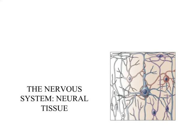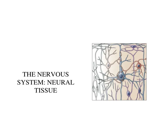
THE NERVOUS SYSTEM: NEURAL TISSUE
510 likes | 1k Views
THE NERVOUS SYSTEM: NEURAL TISSUE. Two organ systems coordinate and direct activities of body. Nervous system Swift, brief responses to stimuli Endocrine system Adjusts metabolic operations Directs long-term changes. An Overview of the Nervous System. Divisions of the nervous system.

THE NERVOUS SYSTEM: NEURAL TISSUE
E N D
Presentation Transcript
Two organ systems coordinate and direct activities of body • Nervous system • Swift, brief responses to stimuli • Endocrine system • Adjusts metabolic operations • Directs long-term changes
Anatomical Classification of the Nervous System • Central Nervous System • Brain and spinal cord • Peripheral Nervous System • All neural tissue outside CNS
Functional divisions of nervous system • Afferent • Sensory information from receptors to CNS • Efferent • Motor commands to muscles and glands • Somatic division • Voluntary control over skeletal muscle • Autonomic division • Involuntary regulation of smooth and cardiac muscle, glands
Cells in Nervous Tissue • Neurons • Neuroglia
Neuroglia (Glia) • about half the volume of cells in the CNS • smaller than neurons • 5 to 50 times more numerous • do NOT generate electrical impulses • divide by mitosis • two types in PNS • Schwann cells • Satellite cells • Four types in the CNS • Astrocytes • Oligodendrocytes • Microglia • Ependymal cells
Astrocytes • Largest of glial cells • Star shaped with many processes projecting from the cell body • Help form and maintain blood-brain barrier • Provide structural support for neurons • Maintain the appropriate chemical environment for generation of nerve impulses/action potentials • Regulate nutrient concentrations for neuron survival • Regulate ion concentrations - generation of action potentials by neurons • Take up excess neurotransmitters • Assist in neuronal migration during brain development • Perform repairs to stabilize tissue
Oligodendrocytes • Most common glial cell type • Each forms myelin sheath around the axons of neurons in CNS • Analogous to Schwann cells of PNS • Form a supportive network around CNS neurons • fewer processes than astrocytes • round or oval cell body
Microglia • few processes • derived from mesodermal cells • that also give rise to monocytes • and macrophages • Small cells found near blood vessels • Phagocytic role - clear away dead cells • protect CNS from disease through phagocytosis of microbes • migrate to areas of injury where they clear away debris of • injured cells - may also kill healthy cells
Ependymal Cells • epithelial cells arranged in a • single layer • range in shape from cuboidal • to columnar • Form epithelial membrane lining cerebral cavities (ventricles) & central canal - that contain CSF • Produce & circulate the cerebrospinal fluid (CSF) found in these chambers • CSF = colourless liquid that protects the brain and SC against • chemical & physical injuries, carries oxygen, glucose and other necessary • chemicals from the blood to neurons and neuroglia
PNS: Satellite Cells • Flat cells surrounding PNS axons • Support neurons in the PNS
PNS: Schwann Cells • each cell surrounds multiple unmyelinated PNS axons with a single layer of its plasma membrane • Each cell produces part of the myelin sheath surrounding an axon in the PNS • contributes regeneration of PNS axons
Neurons • what is the main defining characteristic of neurons? • have the property of electrical excitability - ability to produce • action potentials or impulses in response to stimuli
Representative Neuron http://www.horton.ednet.ns.ca/staff/selig/Activities/nervous/na1.htm • -neurofilaments or neurofibrils give cell shape and support - bundles of • intermediate filaments • -microtubules move material inside cell • -lipofuscin pigment clumps (harmless aging) - yellowish brown • 1. cell body or soma • -single nucleus with prominent nucleolus • -Nissl bodies • -rough ER & free ribosomes for protein synthesis • -proteins then replace neuronal cellular components for growth • and repair of damaged axons in the PNS
Neurons 2. Cell processes = dendrites (little trees) - the receiving or input portion of the neuron -short, tapering and highly branched -surfaces specialized for contact with other neurons -cytoplasm contains Nissl bodies & mitochondria
3. Cell processes = axons • Conduct impulses away from cell body-propagates nerve impulses to another neuron • Long, thin cylindrical process of cell • contains mitochondria, microtubules & neurofibrils - NO ER/NO protein synth. • joins the soma at a cone-shaped elevation = axon hillock • first part of the axon = initial segment • most impulses arise at the junction of the axon hillock and initial segment = trigger zone • cytoplasm = axoplasm • plasma membrane = axolemma • Side branches = collaterals arise from the axon • axon and collaterals end in fine processes called axon terminals • Swollen tips called synaptic end bulbs contain vesicles filled with neurotransmitters
Structural Classification of Neurons • Based on number of processes found on cell body • multipolar = several dendrites & one axon • most common cell type in the brain and SC • bipolar neurons = one main dendrite & one axon • found in retina, inner ear & olfactory • unipolar neurons = one process only, sensory only (touch, stretch) • develops from a bipolar neuron in the embryo - axon and dendrite fuse and then branch into 2 branches near the soma - both have the structure of axons (propagate APs) - the axon that projects toward the periphery = dendrites
Structural Classification of Neurons • Named for histologist that first described them or their appearance • Purkinje = cerebellum • Renshaw = spinal cord • others are named for shapes • e.g. pyramidal cells
Functional Classification of Neurons • Sensory (afferent) neurons • transport sensory information from skin, muscles, joints, sense organs & viscera to CNS • Motor (efferent) neurons • send motor nerve impulses to muscles & glands • Interneurons (association) neurons • connect sensory to motor neurons • 90% of neurons in the CNS
Terms to know • membrane potential = electrical voltage difference measured across the membrane of a cell • resting membrane potential = membrane potential of a neuron measured when it is unstimulated • results from the build-up of negative ions in the cytosol along the inside of the neuron’s PM • the outside of the PM becomes more positive • this difference in charge can be measured as potential energy – measured in millivolts • polarization • depolarization • repolarization • hyperpolarization
The electric potential across an axonal membrane can be measured • the differences in positive and • negative charges in and out • of the neuron can be measured by • electrodes = resting membrane potential • -ranges from -40 to -90 mV
ion channels in the PM of neurons and muscles contributes to their excitability • when open - ions move down their concentration gradients • channels possess gates to open and close them • two types: gated and non-gated Ion Channels • 1. Leakage (non-gated) or Resting channels: are always open, contribute to the resting potential • -nerve cells have more K+ than Na+ leakage channels • -as a result, membrane permeability to K+ is higher • -K+ leaks out of cell - inside becomes more negative • -K+ is then pumped back in • 2. Gated channels: open and close in response to a stimulus • A. voltage-gated: open in response to change in voltage - participate in the AP • B. ligand-gated: open & close in response to particular chemical stimuli (hormone, neurotransmitter, ion) • C. mechanically-gated: open with mechanical stimulation
The resting potential, generated mainly by open “resting”, non-gated K+ channels -the number of K+ channels dramatically outnumbers that of Na+ -however, there are a few Na leak channels along the axonal membrane ECF AXON
Action Potential • Resting membrane potential is -70mV • triggered when the membrane potential reaches a threshold usually -55 MV • if the graded potential change exceeds that of threshold – Action Potential • Depolarization is the change from -70mV to +30 mV • Repolarization is the reversal from +30 mV back to -70 mV) • action potential = nerve impulse • takes place in two stages: depolarizing phase (more positive) and repolarizing phase (more negative - back toward resting potential) • followed by a hyperpolarizing phase or refractory period in which no new AP can be generated http://www.blackwellpublishing.com/matthews/channel.html
The action potential • 1. neuron is at resting membrane potential (resting MP) • 2. neuron receives a signal • Neurotransmitter (NT) • 3. NT binds ligand-gated sodium channel • 4. LGNa channel opens • 5. Na flows into neuron = depolarization • Inside of neuron (i.e. MP) becomes more positive • 6. if neuron depolarizes enough to Threshold = Action Potential (AP) • 7. 1st stage of AP – opening of voltage-gated Na channels • 8. even more Na flows in through VGNa channels = BIG depolarization • Membrane potential goes from negative to positive • 9. closing of VGNa channels & opening of voltage-gated K channels • 10. BIG outflow of potassium through VGK channels = repolarization • Inside of neuron (MP) becomes more negative • 11. neuron repolarizes so much – it goes past resting and hyperpolarizes • 12. closing of VGK channels • 13. all voltage-gated channels closed, Na/K pump “resets” ion distribution to resting situation
Action Potential 9. 8. 10. 6. 11. 13. 12.
Continuous versus Saltatory Conduction • Continuous conduction (unmyelinated fibers) • An action potential spreads (propagates) over the surface of the axolemma • as Na+ flows into the cell during depolarization, the voltage of adjacent areas is effected and their voltage-gated Na+ channels open • step-by-step depolarization of each portion of the length of the axolemma http://highered.mcgraw-hill.com/sites/0072437316/student_view0/chapter45/animations.html#
Saltatory Conduction • Saltatory conduction • -depolarization only at nodes of Ranvier - areas along the axon that are unmyelinated and where there is a high density of voltage-gated ion channels • -current carried by ions flows through extracellular fluid from node to node http://www.blackwellpublishing.com/matthews/actionp.html
Rate of Impulse Conduction • Properties of axon • Presence or absence of myelin sheath • Diameter of axon
Action Potentials in Nerve and Muscle • Entire muscle cell membrane versus only the axon of the neuron is involved • Resting membrane potential • nerve is -70mV • skeletal & cardiac muscle is closer to -90mV • Duration • nerve impulse is 1/2 to 2 msec • muscle action potential lasts 1-5 msec for skeletal & 10-300msec for cardiac & smooth • Fastest nerve conduction velocity is 18 times faster than velocity over skeletal muscle fiber
Synapses Synapse: Site of intercellular communication between 2 neurons or between a neuron and an effector (e.g. muscle – neuromuscular junction) • Permits communication between neurons and other cells • Initiating neuron = presynaptic neuron • Receiving neuron = postsynaptic neuron • You can classify a synapse according to: • 1. the action they produce on the post-synaptic neuron – excitatory or inhibitory • 2. the mode of communication – chemical vs. electrical
Synapses – Excitatory vs. Inhibitory • If the NT depolarizes the postsynaptic neuron = excitatory • The depolarization event is often called an excitatory postsynaptic potential (EPSP) • Opening of sodium channels or other cation channels (inward) • Some NTs will cause hyperpolarization = inhibitory • The hyperpolarization event is often called an inhibitory postsynaptic potential (IPSP) • Opening of chloride channels (inward) or potassium channels (outward) • Neural activity depends on summation of all synaptic activity • Excitatory and inhibitory
Synapses • Electrical • Direct physical contact between cells required • Conducted through gap junctions • Two advantages over chemical synapses • 1. faster communication – almost instantaneous • 2. synchronization between neurons or muscle fibers • e.g. heart beat
Chemical Synapse • Synapse • Most are axodendritic axon -> dendrite • Some are axoaxonic – axon > axon http://www.lifesci.ucsb.edu/~mcdougal/neurobehavior/modules_homework/lect3.dcr
Synapses – Chemical vs. Electrical • Chemical - Most common type of synapse • Membranes of pre and postsynaptic neurons do not touch • Space = Synaptic cleft • the AP cannot travel across the cleft – release of neurotransmitters • The neurotransmitter induces a postsynaptic potential in the PS neuron • if the potential is an EPSP – excitatory and an AP results • If the potential is an IPSP – inhibitory and NO AP results (e.g. glycine or GABA) • Communication in one direction only http://www.blackwellpublishing.com/matthews/nmj.html
The Neuromuscular Junction • end of neuron (synaptic terminal or axon bulb) is in very close association • with the muscle fiber • distance between the bulb and the folded sarcolemma = synaptic cleft • nerve impulse leads to release of neurotransmitter(acetylcholine) • N.T. binds to receptors on myofibril surface • binding leads to influx of sodium, potassium ions (via channels) • eventual release of calcium by sarcoplasmic recticulum = contraction • Acetylcholinesterase breaks down ACh • Limits duration of contraction
The Events in Muscle Contraction • AP generated at trigger zone in • pre-synaptic neuron • 2. AP arrives in end bulb – causes entry • of calcium into end-bulb – release • of Ach • Binding of Ach to ligand-gated Na • channels on muscle PM (Ach receptors) • Na enters muscle cell – depolarization • Muscle membrane potential reaches • threshold = Action Potential • 6. AP travels through PM of muscle cell into • T-tubules • 7. AP “passes by” sarcoplasmic reticulum – • release of calcium into muscle cell • 8. Ca binds troponin-tropomyosin complex & • “shifts” it off myosin binding site • 9. Cross-bridging between actin and myosin, • pivoting of myosin head = Contraction • (ATP dependent)
Neurotransmitters • More than 100 identified • Some bind receptors and cause channels to open • Others bind receptors and result in a second messenger system • Results in either excitation or inhibition of the target • 1. small molecules: Acetylcholine (ACh) • -All neuromuscular junctions use ACh • -ACh also released at chemical synapses in the PNS and by some CNS neurons • -Can be excitatory at some synapses and inhibitory at others • -Inactivated by an enzyme acetylcholinesterase
Neurotransmitters 2. Amino acids: glutamate & aspartate & GABA • Powerful excitatory effects • Glutamate is the main excitatory neurotransmitter in the CNS • Stimulate most excitatory neurons in the CNS (about ½ the neurons in the brain) • Binding of glutamate to receptors opens calcium channels = EPSP • GABA (gamma amino-butyric acid) is an inhibitory neurotransmitter for 1/3 of all brain synapses
Neurotransmitters 3. Biogenic amines: modified amino acids • catecholamines:norepinephrine (NE), epinephrine, dopamine (tyrosine) • serotonin - concentrated in neurons found in the brain region = raphe nucleus • derived from tryptophan • sensory perception, temperature regulation, mood control, appetite, sleep induction • feeling of well being • NE - role in arousal, awakening, deep sleep, regulating mood • epinephrine (adrenaline) - flight or fight response • dopamine - emotional responses and pleasure, decreases skeletal muscle tone
Removal of Neurotransmitter • Enzymatic degradation • acetylcholinesterase • Uptake by neurons or glia cells • neurotransmitter transporters • NE, dopamine, serotonin
Neuropeptides • widespread in both CNS and PNS • excitatory and inhibitory • act as hormones elsewhere in the body • -Substance P -- enhances our perception of pain • -opioid peptides: endorphins - released during stress, exercise • -breaks down bradykinins (pain chemicals), boosts • the immune system and slows the growth of cancer • cells • -binds to mu-opioid receptors • -released by the neurons of the Hypothalamus and by • the cells of the pituitary • enkephalins - analgesics • -breaks down bradykinins (200x stronger than morphine) • -pain-relieving effect by blocking the release of • substance P • dynorphins - regulates pain and emotions • **acupuncture may produce loss of pain sensation because of release of opioid-like substances such as endorphins or dynorphins