Download
1 / 67
670 likes | 752 Views
Explore the neural tissue and functions of the nervous system including organization, cells, and functions like sensory, integrative, and motor capabilities. Learn about neurons, neuroglia, and CNS organization.

E N D
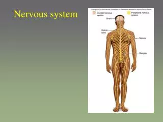
Nervous system includes all neural tissue in body • about 3% of the total body weight • Central Nervous System • Peripheral Nervous System • All neural tissue outside CNS • includes the spinal and cranial nerves
Nervous system functions • 1. sensory function • detect internal and external stimuli • information is sent to CNS • 2. integrative function • integrates = processing of information within the CNS • stores info and also makes decisions once info is processed • processed by interneurons within the CNS
Nervous system functions • 2. integrative function • integrates = processing of information within the CNS • stores info and also makes decisions once info is processed • processed by interneurons within the CNS • 90% of the neurons within the CNS are interneurons
Nervous system functions • 3. motor function • motor commands travel along motor (efferent) neurons within motor nerves • commands are sent to effectors = muscles and glands
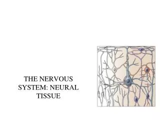
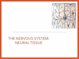
Cells in Nervous Tissue • Neurons • Neuroglia
Neuroglia (Glia) • “glue” • about half the volume of cells in the CNS • 5 to 50 times more numerous • smaller than neurons • do NOT generate electrical impulses • BUT can divide by mitosis • physically support neurons • chemically regulate the neuron’s environment • participate in neural development
Neuroglia (Glia) • Two types in the PNS • Schwann cells • satellite cells • Four types in the CNS • Astrocytes • Oligodendrocytes • Microglia • Ependymal cells
Astrocytes • Largest of glial cells • Most numerous • Star shaped with many processes projecting from the cell body • astrocyte processes make contact with: • the capillaries supplying the CNS • the neurons of the CNS • the pia mater membrane covering the brain and spinal cord
Astrocytes • Functions: • 1. Help form and maintain blood-brain barrier (BBB) • 2. Provide structural support for neurons • 3. Maintain the chemical environment for neurons • 4. Take up excess neurotransmitters • 5. Assist in neuronal migration during brain development • 6. Repair of tissue
Oligodendrocytes • Each forms myelin sheath around the axons of neurons in CNS • Analogous to Schwann cells of PNS • Form a supportive network around CNS neurons • fewer processes than astrocytes • round or oval cell body
Microglia • Small cells found near blood vessels • 15% of the glial cells of the CNS • Phagocytic role - clear away dead cells • protect CNS from disease through phagocytosis of microbes • migrate to areas of injury where they clear away debris of • injured cells - may also kill healthy cells
Ependymal Cells • epithelial cells arranged in a • single layer • range in shape from cuboidal • to columnar • Form epithelial membrane lining cerebral cavities (ventricles) & the spinal cord’s central canal • Produce & circulate the cerebrospinal fluid (CSF) found in these chambers • CSF = colorless liquid that protects the brain and SC against chemical & physical injuries, carries oxygen, glucose and other necessary chemicals from the blood to neurons and neuroglia
PNS: Satellite Cells • Flat cells surrounding PNS cell bodyies • Support neurons in the PNS & help regulate the chemical environment surrounding the neurons • also found in the ganglion of the PNS
PNS: Schwann Cells • each cell surrounds multiple unmyelinated PNS axons with a single layer of its plasma membrane • each cell produces part of the myelin sheath surrounding an axon in the PNS • contributes to regeneration of PNS axons
Neurons • what is the main defining characteristic of neurons? • have the property of electrical excitability - ability to produce • action potentials or impulses in response to stimuli
Representative Neuron http://www.horton.ednet.ns.ca/staff/selig/Activities/nervous/na1.htm • 1. cell body or soma (or perikaryon) • -single nucleus with prominent nucleolus (high synthetic activity) • -neurofilaments give neuron shape and support • -Nissl bodies • -rough ER & free polyribosomes for protein synthesis
Representative Neuron -two kinds of processes (nerve fibers) that emerge from the body of the neuron: dendrites & axons
Dendrites - (little trees) - the receiving or input portion of the neuron -short, tapering and highly branched -surfaces specialized for contact with other neurons
Axons • conduct impulses away from cell body toward synaptic end bulbs • comprised of microtubules & neurofilaments • joins the soma at a cone-shaped elevation = axon hillock • first part of the axon = initial segment • most impulses arise at the junction of the axon hillock and initial segment =trigger zone – site of ion channels
Axons • cytoplasm = axoplasm • plasma membrane = axolemma • side branches = collaterals arise from the axon • axon and collaterals end in fine processes called axon terminals • swollen tips in the terminals = synaptic end bulbs • contain vesicles filled with neurotransmitters for release
Neuron Classification • can classify neurons two main ways: • 1. Structural • 2. Functional
Structural Classification of Neurons • 1. structural: based on number of processes found on cell body • multipolar = several dendrites & one axon • bipolar neurons = one main dendrite & one axon • pseudo-unipolar neurons = one process only
Structural Classification of Neurons • Purkinje = cerebellum • Renshaw = spinal cord • can classify by the name of the histologist that first described them • or can classify them by their appearance
Functional Classification of Neurons • Sensory (afferent) neurons • transport sensory information from skin, muscles, joints, sense organs & viscera to CNS • Motor (efferent) neurons • send motor nerve impulses to muscles & glands • Interneurons (association/integrative) neurons • connect sensory to motor neurons • 90% of neurons in the body
Terms to know • membrane potential= electrical voltage difference measured across the membrane of a cell • resting membrane potential = membrane potential of a neuron measured when it is unstimulated • results from the build-up of negative ions in the cytosol along the inside of the neuron’s plasma membrane (PM) • the outside of the PM becomes more positive • this difference in charge can be measured as potential energy – measured in millivolts
Terms to know • polarization – change in membrane potential away from resting membrane potential (RMP) • depolarization – MP changes from RMP to become more positive • hyperpolarization – MP changes from RMP to become more negative • repolarization – MP “Returns to Resting” MP
Ion Channels • membrane potential changes due to movement of ions in and out of the neuron • ions move through ion channels • when open - ions diffuse down their concentration gradients • two types: gated and non-gated
Non-Gated Ion Channels • 1. Leakage (non-gated) or Resting channels: are always open, contribute to the resting potential • -ions flow down their concentration gradients • -Na+ flows INTO the neuron • -K+ flows OUT OF the neuron • -Na+/K+ pumps returns these ions – against their concentration gradients • -resting membrane potential is established
The resting membrane potential is generated mainly by open “resting”, non-gated K+ channels -the number of K+ leak channels dramatically outnumbers that of Na+ ECF AXON
Non-Gated Ion Channels • 2. Gated channels:channels can possess gates to open and close them • open and close in response to a stimulus
Non-Gated Ion Channels • gated ions channels: • A. ligand-gated: open & close in response to chemical stimuli (hormones, neurotransmitters, ions) • B. voltage-gated: open in response to change in voltage C. mechanically-gated: open with mechanical stimulation
gated channels control the ion permeability of a neuron • changes in channel permeability create electrical signals in a neuron • there are two types of electrical signals • 1. Graded Potentials • 2. Action Potentials
Graded Potentials • reflect the strength of the stimulus that initiates them • large stimulus = strong graded potential • small stimulus = weak graded potential • produce depolarizations or hyperpolarizations • occur in the dendrites or cell bodies • in the CNS and motor neurons - occur upon the opening of ligand-gated ion channels • in sensory neurons – occur upon the opening of mechanically-gated channels • graded potentials are how the neuron achieves threshold
http://www.blackwellpublishing.com/matthews/channel.html Action Potential • action potential = nerve impulse • takes place in three stages: • 1. depolarization phase • 2. repolarizing phase • 3. hyperpolarizing phase or refractory period in which no new AP can be generated
Action Potential • Resting membrane potential is ~-70mV • AP is triggered when the membrane potential reaches and exceeds a threshold (usually -55 MV)
Action Potential: Reaching Threshold • 1. neurotransmitters bind LIGAND-GATED ion channels in the dendrites and the cell body • 2. influx of positive ions into the neuron – usually Na+ • 3. slow depolarization to threshold voltage
Action Potential: Post-Threshold • 1. threshold is reached – opening of VOLTAGE-GATEDNa+ channels & large depolarization results as +ve ions flow into the neuron • 2. VG-Na+ channels close and VG-K+ channel open – at ~+50mV • repolarization as +ve ions flow out of the neuron
Action Potential: Hyperpolarization • 3. VG-K+ channels close over a prolonged duration vs. VG-Na+ • some VG-K+ stay open longer than others • the inside of the neuron drops below RMP = hyperpolarization • refractory period = typical stimulus will not trigger another AP • Na/K pumps “reset” the ions – RMP re-established
hyperpolarization channels are still open. This produces the hyperpolarization phase. As these K+ gates eventually close, resting membrane potential is re-established
Medical Application: Local Anesthetics • Prevent opening of voltage-gated Na+ channels • Nerve impulses cannot pass the anesthetized region • Novocaine and lidocaine – blocks nerve impulses along nerves that detect pain
Continuous versus Saltatory Conduction • Continuous conduction (unmyelinated fibers) • An action potential spreads (propagates) over the surface of the axolemma http://highered.mcgraw-hill.com/sites/0072437316/student_view0/chapter45/animations.html#
Saltatory Conduction • Saltatory conduction • -depolarization only at Nodes of Ranvier - areas along the axon that are unmyelinated and where there is a high density of voltage-gated ion channels • -current carried by ions flowing through extracellular fluid from node to node http://www.blackwellpublishing.com/matthews/actionp.html
Rate of Impulse Conduction • not related to stimulus strength • Properties of axon • Presence or absence of myelin sheath • myelin = 5 to 7X faster • Diameter of axon • larger = faster conduction
Action Potentials in Nerve and Muscle • another excitable cell type = muscle • muscle cells can generate their own action potential in response to stimulation by the neuron • some notable differences between Muscles vs. Neurons: • 1. entire muscle cell membrane is involved versus only the axon of the neuron • 2. resting membrane potential differ • nerve is -70mV • skeletal & cardiac muscle is closer to -90mV • 3. AP duration is longer in muscles • nerve impulse is 1/2 to 2 msec • muscle action potential lasts 1-5 msec for skeletal & 10-300msec for cardiac & smooth • BUT fastest nerve conduction velocity is 18 times faster than velocity over skeletal muscle fiber
Synapse • Synapse = Site of intercellular communication between two neurons OR between a neuron and an effector (e.g. muscle) • Permits communication between neurons and other cells • Initiating neuron = presynaptic neuron • Receiving cell = postsynaptic cell • Most synapses are axodendritic axon -> dendrite • Some synapses are axoaxonic – axon > axon