Download
1 / 20
210 likes | 405 Views
This assessment explores the complexities of the cardiac cycle, detailing events such as atrial and ventricular systole and diastole, as well as the associated heart sounds. It analyses pressure and volume changes in the left atrium, left ventricle, and aorta throughout the cardiac cycle. Key mechanisms, including the roles of the sinoatrial and atrioventricular nodes, are outlined. Additionally, it addresses atherosclerosis, coronary thrombosis, and factors influencing the incidence of coronary heart disease, emphasizing their impact on cardiovascular health.

E N D
Assessment Statements • H.5.1 Explain the events of the cardiac cycle, including atrial and ventricular systole and diastole, and heart sounds. • H.5.2 Analyse data showing pressure and volume changes in the left atrium, left ventricle and the aorta, during the cardiac cycle. • H.5.3 Outline the mechanisms that control the heartbeat, including the roles of the SA (sinoatrial) node, AV (atrioventricular) node and conducting fibres in the ventricular walls. • H.5.4 Outline atherosclerosis and the causes of coronary thrombosis. • H.5.5 Discuss factors that affect the incidence of coronary heart disease.
Events of the cardiac cycle • Sinoatrial (SA) node fires electrical signal throughout walls of atria to begin the cardiac cycle • the SA signal causes atria to undergo systole - atria contract & blood flows to the ventricles • SA signal reaches atrioventricular (AV) node, which spreads signal throughout the Purkinje fibres • causing ventricles to undergo systole, • when ventricular pressure rise above atrial pressure, atrioventricular valves slap shut, causing “lub” sound • semi-lunar valves open, blood is pumped into arteries • as ventricles relax, ventricular pressure falls, pressure in artery exceeds pressure in ventricle, semilunar valves close, causing “dub” sound • atrioventricular valves open, ventricles begin diastole and start filling with blood • all four chambers are in diastole and filling with blood • when atria are completely filled with blood and ventricles are 70 % full, the cycle ends & the cycle repeats
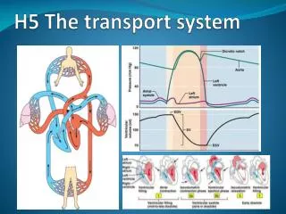
Pressure & volume changes in the left atrium, left ventricle &the aorta, during the cardiac cycle
Pressure & volume changes in the left atrium, left ventricle & the aorta, during the cardiac cycle
Mechanisms that control the heartbeat • heartbeat is myogenic i.e. initiated in heart muscle itself • SA node (pacemaker) sends waves of excitation to atria • stimulus is then passed to the AV node • conducting fibres (Purkinje fibres) conducts impulses to lower ventricles • heartbeat is moderated by ANS i.e. parasympatheticnervous system
Role of the (SA) sinoatrial node in the cardiac cycle • SA node is located in the wall of right atrium of heart muscle • SA has characteristics of both nerve and muscle tissue • SA node initiates each impulse & acts as pacemaker of the heart • no nerve impulses needed for contraction i.e. SA is myogenic • SA is connected to nerves which slow or accelerate heart rate; • impulses spread out in all directions through walls of atria stimulating atrial systole (contraction) • fibres in walls of atria prevent impulses from reaching ventricles • impulses only reach AV node after atrial contraction
Role of the AV (atrioventricular) in the cardiac cycle • AV node situated at the base of the right atrium, it receives impulse (wave of excitation) from atrial walls • AV node causes time delay before the impulse is passed to the ventricular tissue • AV node then passes the impulse to modified muscle fibres called Purkinje fibres (bundle of His) in the ventricular wall
Role of conducting fibres in the cardiac cycle • AV bundles (bundle of His) originates from AV node, run along interventricular septum & branch into Purkinje fibres • AV bundles conducts nerve impulse from AV node to Purkinje fibres • Purkinje fibres are specialized muscle fibres found in ventricular muscles • Purkinje fibres are insulated from the muscle and do not cause contraction • their function is to relay impulses from the AV bundle to the ventricle muscles causing a contraction • the impulse emerges into the muscle at the apex of the heart so that the ventricular contraction begins at the apex spreading upwards
Atherosclerosis and coronary thrombosis • Atherosclerosis is a progressive degeneration of artery walls • lipids (cholesterol) are deposited on endothelium narrowing the artery lumen • fibrous tissue may also be laid down impeding blood flow, causing platelets to stick together • clotting factors may then be released, a blood clot within the vessel or thrombus may then form • if atherosclerosis occurs in coronary artery, coronary thrombosis, flow of blood to part of heart muscle is reduced or stopped leading to lack of glucose & oxygen • myocardial infarction, heart attack, cardiac arrest, heart failure may result
Causes of coronary thrombosis • atheroma, fatty deposits in arteries occurs • atheroma causes hardening of arteries i.e. atherosclerosis (arteriosclerosis) • rough surface in artery lumen causes rupture of platelets • blood clots form in coronary artery - coronary thrombosis
Incidences of coronary heart disease events • Incidences of coronary heart disease events was significantly associated with: with age; gender (men); diabetes & hyperlipidemia i.e. high lipid levels usually cholesterol • Is there significant difference between events of gender (men), diabetes & hyperlipidemia? • Support your answer. • * BNP = brain natriuretic peptide
Factors that affect the incidence of coronary heart disease • genetic factors – some people are predisposed for high cholesterol levels & high blood pressure • age – older people are at greater risk due to less elasticity in arteries • sex – males are at greater risk of heart disease than pre-menopausal women because they have less estrogen, as estrogen protects against heart disease • smoking – nicotine causes vasoconstriction of blood vessels, increases blood pressure, heart-rate & decreases oxygenation of heart muscle • diet – eating too much saturated fat, high cholesterol & LDL in blood leads to plaque formation in arteries - coronary thrombosis • exercise – helps reduce high blood pressure, reduces the rate of fatty deposits building up in the inner lining of arteries & thickens the heart muscle walls so they pump blood more efficiently • obesity – lead to increase in blood pressure & leads to plaque formation in arteries • highsalt diet , excessive amounts of alcohol& stresscan also affect the incidences of coronary heart disease
Revision Questions • Explain the events of the cardiac cycle. [7] • Describe the mechanisms that control the heartbeat. [4] • Outline how the contraction of the atria and the ventricles is controlled. [4] • Explain the role of the SA (sinoatrial) node in the cardiac cycle. [6] • Explain the role of the AV (atriolventricular) node in the cardiac cycle. [4] • Explain the role of the conducting fibres in the cardiac cycle. [4] • Outline the condition atherosclerosis and how it may cause coronary thrombosis. [5] • Outline how coronary thrombosis can be caused.[3] • Discuss the factors which that affect the incidence of coronary heart disease. [7]
The graph above shows pressure changes in the left atrium, left ventricle and the aorta, during the cardiac cycle. Explain the changes in pressure.
The graph above shows volume changes in the left ventricle during the cardiac cycle. Explain the changes in volume.