Download
1 / 65
740 likes | 1.16k Views
The Transport System IB DP Biology: Core + Further Human Physiology (Option H5). Heart Diagrams. Draw five heart diagrams just like the picture above. Show the chambers of the heart. Areas of the body blood goes to or returns from. Show the major blood vessels of the heart.

E N D
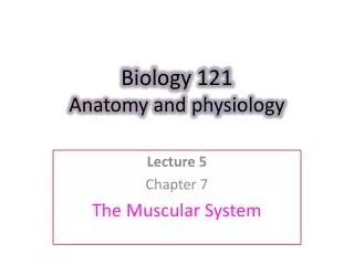
The Transport SystemIB DP Biology: Core + Further Human Physiology (Option H5)
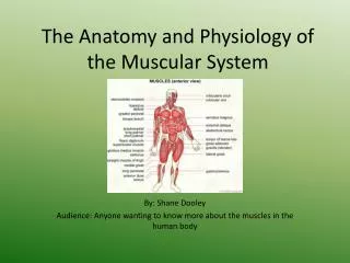
Heart Diagrams • Draw five heart diagrams just like the picture above. • Show the chambers of the heart. • Areas of the body blood goes to or returns from. • Show the major blood vessels of the heart. • Show the valves (atrio-ventricular & semi-lunar). • Electricity of the heart and hormone control.
Heart Diagram • You must be able to diagram the heart including: • Show the four chambers • Show associated blood vessels. • Show the valves • Show the route of blood through the heart.
3 Main Parts of Transport System • Heart – a pump that applies pressure to the blood establishing a pressure gradient. • Blood vessels – passageways through which blood travels & is distributed. • Blood – transport medium within which materials are dissolved or suspended.
What is the transport system transporting? • Oxygen • Nutrients • Antibodies • Hormones • Heat • Carbon dioxide • Urea This transport by the beating heart pumping blood through a network of capillaries, arteries and veins. http://www.beatmap.net
Blood components: • Plasma – dissolves or carries all other components of blood • Erythrocytes(Red blood cells)– transport oxygen in hemoglobin molecules • Leucocytes(White blood cells) • Phagocytes: ‘eat up’ pathogens and dead cells • Lymphocytes(B-cells, T-cells) for immune response • Platelets– clotting of blood following damage to cells or erythrocytes http://www.denniskunkel.com/
Introduction to the heart • Your heart is a specialized pump made of muscle and its job is to circulate blood through your body effectively. • On average your heats beats 60 – 100 times per minute, every single minute of the day. Because your heart never gets to rest it is made of a special type of muscle called cardiac muscle that does not tire easily.
A The heart What is this? G R I G H T H L E F T I C J F K L E D B Thick cardiac muscle
Coronary arteries These arteries supply oxygen & nutrients to heart muscle. A blockage here leads to a heart attack (myocardial infarction. A section of heart muscle does not get enough energy or nutrients and dies.
Virtual heart dissection http://www.gwc.maricopa.edu/class/bio202/cyberheart/anthrt.htm Roll over to see the structures http://www.nucleusinc.com/animation2.php
Action of the heart University of Kentucky – Blood flow Blood enters the heart via veins into the atria Contraction pushed blood into the ventricles Further contraction pushes blood out of the heart into the arteries to be carried to the lungs or body. Atrio-ventricular (Tricuspid & bicuspid (mitral) valves and semi-lunar (pulmonary and aortic) valves prevent backflow of blood Ventricles have higher pressure as they push blood under pressure into the arteries. www.medmovie.com Flow of blood is by a double circuit system – systemic & pulmonary
Double circulation – blood passes through the heart twice on two separate circuits. Deoxygenated blood (low O2, high CO2) returns to the heart via the right atrium. It is pumped from the right atrium to the lungs where CO2 is removed and O2 is picked up. It is now oxygenated blood (High O2 and low CO2 Oxygenated blood enters the left atrium and is pumped from the left ventricle to the body, where oxygen is used for respiration and CO2 is collected as a waste product. It is now deoxygenated & makers its way back to the right atrium and the cycle continues. http://www.kscience.co.uk/animations/blood_system.swf
Concept 42.2: Coordinated cycles of heart contraction drive double circulation in mammals • The mammalian cardiovascular system meets the body’s continuous demand for O2 • Blood begins its flow with the right ventricle pumping blood to the lungs • In the lungs, the blood loads O2 and unloads CO2
Oxygen-rich blood from the lungs enters the heart at the left atrium and is pumped through the aorta to the body tissues by the left ventricle • The aorta provides blood to the heart through the coronary arteries
Blood returns to the heart through the superior vena cava (blood from head, neck, and forelimbs) and inferior vena cava (blood from trunk and hind limbs) • The superior vena cava and inferior vena cava flow into the right atrium
Superior vena cava Capillaries of head and forelimbs Figure 42.6 Pulmonary artery Pulmonary artery Capillaries of right lung Capillaries of left lung Aorta Pulmonary vein Pulmonary vein Left atrium Right atrium Left ventricle Right ventricle Aorta Inferior vena cava Capillaries of abdominal organs and hind limbs
The Mammalian Heart: A Closer Look • The heart contracts and relaxes in a rhythmic cycle called the cardiac cycle • The contraction, or pumping, phase is called systole • The relaxation, or filling, phase is called diastole
Aorta Pulmonary artery Figure 42.7 Pulmonary artery Right atrium Left atrium Semilunar valve Semilunar valve Atrioventricular valve Atrioventricular valve Right ventricle Left ventricle
The heart rate, also called the pulse, is the number of beats per minute • The stroke volume is the amount of blood pumped in a single contraction • The cardiac output is the volume of blood pumped into the systemic circulation per minute and depends on both the heart rate and stroke volume
Four valves prevent backflow of blood in the heart • The atrioventricular (AV) valves separate each atrium and ventricle • The semilunar valves control blood flow to the aorta and the pulmonary artery
The “lub-dup” sound of a heart beat is caused by the recoil of blood against the AV valves (lub) then against the semilunar (dup) valves • Backflow of blood through a defective valve causes a heart murmur
Blood Vessels – Structure & Function • Arteries • Carry blood away from the heart to tissues • Have thick walls to withstand high pressure • Have muscle fibers to help pump blood and even out blood flow. • Have elastic fibers to allow artery wall to stretch & recoil.
Blood Vessels – Structure & Function • Veins • Carry blood back to the heart from tissues. • Have thinner walls because pressure is low. This allows them to be squeezed by adjacent muscles • Have fewer muscles & elastic fibers because pressure is low. • Veins have valves to prevent back flow.
Blood Vessels – Structure & Function • Capillaries • Allow exchange of O2, CO2, nutrients, wastes from tissues/cells. • Have thin wall to allow for rapid diffusion. • Have porous walls to allow phagocytes & tissue fluid to leave • Are narrow so can penetrate all parts of tissues • Have bigger total surface area.
Control of heart beat • A beating heart is due to myogenic muscle contraction. • The sinoatrial (SA) node controls heart beat (pacemaker) • A wave of excitation is sent from the SA node causing the atria to contract. • This excitations is conducted to the atrioventricular (AV) node where it is passed through nerves to the muscles of the ventricles causing them to contract. http://www.emedicinehealth.com Myogenic initiation of the contraction means that the heart does not stop beating. It is unique because it initiates its own action potentials!
Normal Rate of Action Potential Discharge • heart cells with the fastest rate of action potential initiation are in the SA node. So the SA node drives the heart & is known as the pacemaker of the heart.
Control of heart beat • Heart rate can be controlled by the autonomic nervous system. The part of the nervous system that responds automatically to changes in body conditions. • The normal heart rate may need to speed up or slow down. • When exercising more CO2 is detected in the blood by chemoreceptor's in the medulla oblongata. A nerve signal is sent to the SA node to speed up HR. • When CO2 levels fall, another nerve (Vagus) reduces HR. • The hormone adrenalin causes a rapid increase in HR in flight-or-flight responses http://medmovie.com Can you convert the control of the heart beat into a flow chart? How is it an example of negative feedback?
Nervous System Central NS Peripheral NS Brain Spinal chord Sensory Division Motor Division Autonomic NS (Involuntary) Somatic NS (Voluntary) Sympathetic (activities that increase energy consumption- flight or fight) Parasympathetic (Normalizes body functions) • Remember this?
Heart rate is moderated by the ANS. • The parasympathetic nervous system decreases heart rate by decreasing the AV nodes excitability thru the vagus nerve. • Sympathetic stimulation on the SA node increases its rate of depolarization so that threshold is reached more rapidly. HR increases • Sympathetic nerve endings release epinephrine (adrenaline).
Echocardiograms (ECG) measure heart rate Use the animation and the graph to explain what is happening at the various stages of the heart beat. At each stage: What muscles are contracting? Which are relaxed? What valves are open? Which are closed? Explain: Why is the muscle of the left ventricle thicker than on the right? Further reading (HL Option H) What causes heart sounds?
Transport SystemIB DP Biology Further Human Physiology Option H5
A The heart – a review What is this? G R I G H T H L E F T I C J F K L E D B Thick cardiac muscle
Events of the Cardiac Cycle • The cardiac cycle consists of alternate periods of systole (contraction and emptying) & diastole (relaxation and filling). • SA (sino atrial) node (pacemaker) receives signal to fire when ventricles 70% full (late in ventricular diastole). • AV (atrio ventricular) valve opens and blood fills ventricle to maximum (atrial systole).
Pressure increase in ventricle closes AV valve (ventricular systole). • AV node fires • Purkinje fibers carry impulses to all areas of ventricles for simultaneous firing • Pressure increase causes semilunar valve to open • Blood pumped from ventricle to aorta (systole sound) & ventricular diastole • Pressure lowers in ventricle closing semilunar valve (diastole sound)
Pressure in ventricle lower than atria so AV valve opens. • Increases blood ventricular volume • Both atria & ventricles relaxed (diastole) • Atria receives blood from veins • Cycle repeats
Heart sounds • The first heart sound is associated with closure of the AV valves. • The second sound is associated with closure of the semilunar valves. • Opening valves does not produce any sound • Sounds are caused by vibrations set up within the walls of the ventricles and major arteries. Not the valves snapping shut.
Atrial and ventricular diastole Figure 42.8-1 1 0.4 sec
Atrial and Atrial systole and ventricular ventricular diastole diastole 2 Figure 42.8-2 1 0.1 sec 0.4 sec
Atrial and Atrial systole and ventricular Ventricular systole and atrial ventricular diastole diastole diastole 2 Figure 42.8-3 1 0.1 sec 0.3 sec 0.4 sec 3
The cardiac cycle – click on the tutorial for a description of what happens at each stage. Try to follow the actions of the heart on the graph.
Electrical Activity of the Heart(Mechanisms that control heartbeat) • Cardiac muscle cell contraction is triggered by action potentials sweeping across the muscle cell membranes. • The heart generates it own action potentials a property known as autorhythmicity. • 99% of cardiac muscle cells are contractile cells which do the mechanical work of pumping. • The reminder of cardiac cells are autorhythmic (self-excitable) cells which initiate & conduct action potentials.
Pacemaker activity – autorhythmic cell membrane potential slowly depolarizes, or drifts, between action potentials until threshold is reached then Pacemaker sends waves of excitation.
Control of heart beat Myocardium = heart muscle Myogenic originates in the heart SA node generate impulse independently or it can be moderated by the ANS. Signal causes contraction of the atrium Signal travels through conducting fibers to AV node - there is a delay there Signal carried by conducting fibers to lower ventricles which contract. http://www.webmd.com/heart/conduction-system-of-the-heart http://medmovie.com/mmdatabase/mediaplayer.aspx?message=VG9waWNpZD0wJkNsaWVudElEPTQ0-d7XJsZJWVDg%3d
Maintaining the Heart’s Rhythmic Beat • Some cardiac muscle cells are self-excitable, meaning they contract without any signal from the nervous system • The sinoatrial (SA) node, or pacemaker, sets the rate and timing at which cardiac muscle cells contract • Impulses that travel during the cardiac cycle can be recorded as an electrocardiogram (ECG or EKG)
Figure 42.9-1 1 SA node (pacemaker) ECG