Chromosome Dropping
This study evaluates various chromosome spreading techniques using the murine B16 melanoma cell line to identify the optimal method for visualizing metaphase chromosomes. Chromosome abnormalities are critical in melanoma research, making karyotype analysis essential. Two primary techniques tested include varying the dropping height and adjusting relative humidity during the spreading process. Findings indicate challenges faced, as both techniques resulted in non-visualization of chromosomes, highlighting the need for refined methods in karyotyping to achieve successful chromosome spreads.

Chromosome Dropping
E N D
Presentation Transcript
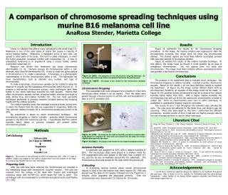
A comparison of chromosome spreading techniques using murine B16 melanoma cell lineAnaRosa Stender, Marietta College Introduction Results Cancer is a disease that affects many individuals in the world today [1]. Melanoma is one of the rarer types of cancer but causes a majority of cancer leading deaths. Melanoma, a malignant tumor in skin cells, can spread to other parts of the body. This term is called metastasis, in which the tumor possesses increased motility and invasiveness [2]. A way to understand melanoma is to experiment using a mouse model, namely murine B16 melanoma [5]. Research has shown that melanoma can cause chromosome abnormalities [3, 4]. Humans possess 46 chromosomes within a cell; whereas, mice have 40 chromosomes [6]. A way to study the abnormalities in chromosomes is to create a karyotype. A karyotype is a photographic representation of all the chromosomes within a cell. The karyotype can show characteristics, such as absolute size, number, and type of chromosomes. To make a karyotype, chromosome spreads can be created to allow the observer to visually see the metaphase chromosomes within the cell [7]. To prepare a well-spread chromosome picture, many techniques have been tested to see which technique presents the best spreads. Factors that affect chromosome spreads include: dropping height, wetness and angle of slide, drying time, and relative humidity [8]. The two most successful techniques performed in previous research included altering the dropping height and the relative humidity. The relative humidity factor has emphasis towards a faster drying time, in which the fixative that the cells are suspended in evaporates faster [9]. The optimum relative humidity for a chromosome spread was proven to be 55%. My experiment is based on which chromosome technique - the chromosome dropping or relative humidity - presents better chromosome spreads in the B16 F10 melanoma cell line. I hypothesize that the relative humidity factor, based on previous research, will present better chromosome spreads. Figure 2b represents the results for the chromosome dropping procedure. In this image, the Giemsa solution was supposed to stain the chromosomes; however, this image does not show any chromosomes stained. The stained regions are most likely leftover associated proteins that were also stained by the Giemsa solution. Figure 3b exhibits the results of the relative humidity technique. At first glance, this image looks as though it could possibly be an area of metaphase chromosomes. Yet, the picture does not show any chromosomes. The image most likely contains leftover associated proteins and possibly a few broken chromosomes. The purpose of my experiment was to evaluate which technique - the chromosome dropping or relative humidity - resulted in better chromosome spreads. Based on my results, I do not have definitive results to support my hypothesis. In Figure 2b, the image reveals leftover debris with no chromosomes; therefore, an analysis of this image could not be made. In Figure 3b, the possible broken chromosomes may be a result of the relative humidity being higher than 55%. With a higher relative humidity, the chromosomes may spill out of the cell causing the chromosomes to break or scatter [8]. Since no chromosomes were seen in either technique, no qualitative or quantitative analysis could be concluded. One source of error I had throughout the semester was culturing the cells. The cells were not adhering to the flask; thus, cell division was not occurring. Most cells were dead by the time I performed the different procedures. Another source of error I had was not having enough cells by the end of preparing them to use for either technique. [1] Wingo PA, Tong T, Bolden S. 1995. Cancer Statistics. Journal of Clinical CA Cancer 45: 8-30. [2] Nowell PC. 1986. Mechanism of tumor progression. Cancer Research 46: 2203-2207. [3] Frohling S, Dohner H. 2008. Chromosome abnormalities in cancer. New England Journal of Medicine 349 (7): 722-734. [4]Robertson GP, Coleman AB, Lugo TG. 1996. Mechanism of Human Melanoma Cell Growth and Tumor Suppression by Chromosome 61. Cancer Research 56: 1635-1641. [5]Slovak ML, Hoeltge GA, and Ganapathi R. 1986. Abnormally banded chromsomal regions in doxorubicin-resistant B16-BL6 murine melanoma cells. Cancer Research 46: 4171-4177. [6]Nesbitt MN, Francke U. 1973. A system of nomenclature for band patterns of mouse chromosomes. Chromosoma 41: 145-158. [7]Qu YY, Xing LY, Hughes ED, Saunders TL. 2008. Chromosome dropper tool: effect of slide angles on chromosome spread quality for murine embryonic stem cells. Journal of Histotechnology 31 (2): 75-79. [8] Deng W, Tsao S, Lucas J, Leung CS, Cheung A. 2002. A new method for improving metaphase chromosome spreading. Cytometry Part A 51A: 46-51. [9] Spurbeck JL, Zinsmeister AR, Meyer KJ, Jalal SM. 1996. Dynamics of chromosome spreading. American Journal of Medical Genetics 61: 387-393. [10] Funasaka Y, Mishima Y, Ichihashi M, Sugiyama. 1988. Comparative analysis of oncogene expression and chromosome abnormalities between metastatic and nonmetastatic B16 melanoma clones. Dermatologica 177: 200-211. I would like to thank my capstone advisor Dr. Spilatro for his feedback throughout this past year. Also, a thank you to Dr. Lustofin, Dr. McShaffrey and the entire Biology Department. Lastly, thanks to my family, fellow capstone students, and friends who have always supported me. Conclusions Figure 2a (left): The apparatus for the chromosome dropping technique. An ice bucket with microscope slides was placed 4 feet below the pasteur pipet. Figure 2b (right): The image of the results for the chromosome dropping technique. Chromosome Dropping The suspended cells were dropped from a height of 4 feet onto microscope slides chilled in an ice bucket. Then the slides were passed through a Bunsen burner to set the cells and incubated for 3 days in a 37°C incubator [10]. Relative Humidity A waterbath was prepared at 50°C with a relative humidity of 55%. The microscope slides were placed in a shallow metal lid. The resuspended cells were placed on the slide, from a height of 1 cm, and the lid was immediately placed into the waterbath. The chromosomes dried and spread on the slide within 90 seconds [8]. G-Banding In G-banding, Giemsa solution allows certain chromosomal regions to bind to the dye heavier than other regions [9]. PBS was poured over the slide for 10 minutes, followed by 0.2% Trypsin for 2 minutes, which degraded the associated proteins. Then the chromosomes were stained with 2% Giemsa for 10 minutes. Literature Cited Methods Figure 3a (left): The setup for the relative humidity procedure involved slides being placed in a metal jar lid. Once cells were placed onto the slide, the lid was slightly submerged into the water. Figure 3b (right): The image of the results for the relative humidity technique. Cell Culturing Culture cells (4-7 days in CDMEM) Trypsinize Transfer 1:4 Figure 1: The process of cell culturing for B16 F10 melanoma cells. After 4-7 days in CDMEM, Trypsin was added to the cells to detach them from the bottom of the flask. The volume was divided into four new T25 flasks. Preparing Cells After the cells have bene cultured, the chromosomes are arrested in the metaphase stage with Demecolcine (1ug/ml) solution. The cells are released from the surface of the flask with Trypsin and centrifuged numerous times with 0.075M KCl, which cause the cells to swell. The centrifuged cells are suspended in a methanol and glacial acetic acid fixative (3:1) [10]. Acknowledgements