Download
1 / 65
670 likes | 936 Views
THE ROAD MAP: Today- 4/24: Digestive System & Immune System (4) 4/25, 4/26, 4/29: Consultations (Image data review) 4/26, 4/29, 5/1: Group In-class Presentations Lab Practical: 4/23 and 4/24 Final Exam: 10 :30 AM May 7 th , 2013 ISB 221

E N D
THE ROAD MAP: Today- 4/24: Digestive System & Immune System (4) 4/25, 4/26, 4/29: Consultations (Image data review) 4/26, 4/29, 5/1: Group In-class Presentations Lab Practical: 4/23 and 4/24 Final Exam: 10:30 AM May 7th, 2013 ISB 221 Individual Project Written Papers: dueFriday, 5/3
The Group Project ~10 minutes/presentation 5‐7 slides: We are all about images‐ no words needed! Organization and clarity are importantso REHEARSE! PRESENTATION GOALS: Identify yourselves. Introduce the histological features of your organ using your paraffin images. Orient the audience: planes of section etc. Introduce the markers you used and your predictions. Use your images to demonstrate what stained. Discuss your results including problems or artifactual staining.
Anxieties Are Us: The Group Project • Hands-on opportunities • Not Cookbook! No guarantees. • Beauty is hard to achieve. • Some folks are just plain lucky. • Life is not fair. Get used to it. • Relax! Do your best. Work hard. • Tell the story- but briefly! • Accept responsibility. • Enjoy the ride.
Possible Artifacts FREEZE/THAW DAMAGE KNIFE MARKS OTHERS: Uneven Staining Folded/wrinkled section Particulate debris Antibody nonspecific staining
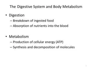
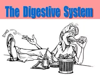
Enteroendocrine Cells • Hormonal release and nerve activity required for coordinating ingestion with secretion and motility • With ingestion of food: • Release of saliva • Release of digestive enzymes • Release of HCl • Release of bile from gall bladder • Motility of gastrointestinal tract • What signals might trigger release of hormones and • digestive enzymes?
Stomach enteroendocrine cells release Gastrin: release from G cells is stimulated by: 1) peptides and amino acids in stomach lumen 2) distention of stomach wall 3) sensory inputs --> neural innervation (GRP) - Parietal cells have gastrin receptors
Small intestine enteroendocrine cells release Choleocystokinin(CCK): CCK released is stimulated by: presence of H+, amino acids, and fatty acids - Pancreatic and gall bladder cells CCK receptors • Release of bile from gall bladder into duodenum • - Release of digestive enzymes from pancreas
PANCREAS: Endocrine / Exocrine Gland http://www.uwgi.org/gut/stomach_03.asp
PANCREAS: Endocrine Gland Islet of Langerhans: most numerous tail of pancreas, 2% volume
Pancreas: Endocrine Function Cells: secretion of insulin in response to low blood glucose, causes uptake and storage of glucose into liver and skeletal muscle; diabetes mellitus Type I and II Cells: secretion of glucagon in response to high blood glucose, causes synthesis of glucose and breakdown of glycogen in liver Cells: secretion of somatostatin; inhibits insulin and glucagon release
GALL BLADDER - lumen lined by simple columnar epithelium - microvilli, tight junctions, mitochondria - smooth muscle - concentrates bile (Na+ actively pumped out of lumen in exchange for H+, water follows) - stores bile until release
Choleocystokinin (CCK) -Gall Bladder smooth muscle cells have CCK receptors CCK RELEASE (INTESTINAL ENDOENDOCRINE CELLS) GALL BLADDER CONTRACTION RELEASE OF BILE INTO LUMEN OF SMALL INESTINE FACILITATED DIGESTION OF LIPIDS
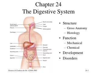
LIVER Right side, beneath diaphragm Lobular “Hepatic” Functions: • Produce of bile • Process and store of nutrients • Phagocytose debris and bacteria • Synthesize blood proteins and coagulation factors • Detoxify & inactivate drugs and toxic substances • Stores, modifies Vitamins A, D, K • Participate in iron metabolism • Modify hormones (thyroxine, growth hormone etc)
Blood Supply: Hepatic Portal System TO LIVER: PORTAL VEIN: oxygen poor blood; from stomach, pancreas, spleen, & intestines. HEPATIC ARTERY: brings oxygen rich blood IN LIVER: HEPATIC SINUSOIDS (2nd capillary bed-portal system) CENTRAL VEIN HEPATIC VEIN VENA CAVA
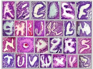
LIVER: Classic Hepatic Lobule Hexagonal; connective tissue septa Bounded by Portal Triads: 1) a branch of the hepatic artery 2) a branch of the portal vein 3) a bile ductule 4) lymphatic vessel (not always present) Central vein (terminal hepatic venule) in center
LIVER: Classic Hepatic Lobule bounded bt portal triads
LIVER: Hepatocytes- arranged in cords; separated by sinusoids • Hepatocyte: 60% of all liver cells • -polyhedral cells • -microvilli: Space of Disse& bile canaliculi. • - extensive ER • abundant mitochondria • - storage granules • of glycogen Perisinusoidal Space SPACE OF DISSE
LIVER: SINUSOIDS Discontinuous capillaries; Squamous endothelial cells Little or no basal lamina; Space of Disse
LIVER: SINUSOIDS Kupffer Cells: protrude into sinusoids; contribute to endothelial lining; macrophages-like; remove blood cells, bacteria Kupffer cells labeled with India Ink
LIVER: SINUSOIDS Ito Cells (hepatic stellate cell): in perisinusoidal space stores Vitamin A-> retinol to form photopigment (rhodopsin) in eye respond to liver inflammation and injury--> scar NORMAL LIVER LIVER INJURY
LIVER: LIVER ACINUS Liver Acinus: Correlates with blood perfusion, metabolic activity and liver pathology; oriented around afferent vascular system
LIVER: LIVER ACINUS Zones correspond to distance from blood supply Zone 1: closest to arterioles, highest oxygen, 1st toxin exposure, necosis due to toxins, first to regenerate Zone 3: farthest from arterioles, poorest supply of oxygen, necrosis due to ischemia
LIVER: BILE PRODUCTION Bile Canaliculus: channel between adjoining hepatocytes carries bile to bile duct
LIVER: BILE PRODUCTION Flow of bile is opposite that of blood flow
LIVER: PORTAL LOBULE Portal Lobule: Region of bile production drained by axial bile duct
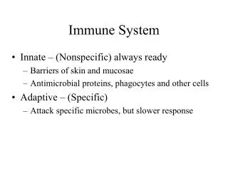
Lymphatic System • monitor body surfaces and internal fluids for antigens and materials for removal • react to potentially harmful substances
Lymphatic Tissues - Concentrated near respiratory tract digestive tract reproductive tract Antigen: foreign substance
Nonspecific Inflammatory Response Mast cells -Histamine release -Vasodilation (increase capillary permeability) Neutrophils -enzymatic digestion of virus Macrophages -cytokine release -phagocytosis -antigen presentation
Specific Responses to Antigen Exposure: Antigen-Presenting Cells (APCs): -APCs include: macrophages and B-lymphocytes 1) endocytoseand degrade antigens 2) display a portion of the antigen coupled to Major Histocompatibility Complex (MHC) II molecules and on cell surface 3) activate other immune cells Humoral Immunity Cell-mediated Immunity Antibody Production Killing of Infected Cells
HumoralImmunity: Antibody Production • Stimulation of B Lymphocytes (B cells) • Antigen processing & presentation: B cells (MHC II) • Stimulation of CD4 Helper T lymphocytes • Production of Interleukins --> stimulate B cells • B cell Proliferation --> Plasma cells & Memory cells
HumoralImmunity: Antibody Production Interleukins (ILs) See also Figure 14.6 in Textbook re: CD40 and CD40L
HumoralImmunity: Antibody Production B-lymphocytes differentiate to form: • Plasma Cells: produce antibodies against a single epitope 1st exposure--> IgM 2nd exposure--> IgG • Memory cells: circulate for future encounters with antigen
With Second Exposure to Antigen: Activation of Memory Cells specific for Antigen Differentiate into Plasma Cells: produce IgGantibody With Secondary Exposure to Antigen: immune response is faster more antibody is produced more rapidly antibodies bind antigen more strongly
Cell-Mediated Immunity Cytotoxic (CD8+) T-lymphocytes attack and destroy virus-infected cells Macrophage engulfs, processes & presents antigen Stimulation of CD4 Stimulation of CD8 Helper T Lymphocytes Cytotoxic T Lymphocytes MHC II MHC I Proliferation Killing of INFECTED cells Release of Interleukin 2
Cell-Mediated Immunity:Cytotoxic CD8 T-lymphocytes granzymes 1)Macrophage presents antigen to CD4+ and CD8+ T cells 2)IL2 stimulates CD4+ and CD8+ proliferation 3)CD8+ T cells recognize virus-infected cells (MHC I) 4)CD8+ release granzymesplus 5)Infected cells die by apoptosis
Cytotoxic T Lymphocytes: • - bind to target cell • exocytosis of granules of perforinand granzymes • Perforins insert into target cell membrane • Granzymes, serine proteases, • enter cell and cause cell death (apoptosis ) via activation of precursors of caspases. http://sprojects.mmi.mcgill.ca/immunology/Perforin.JPG http://users.rcn.com/jkimball.ma.ultranet/BiologyPages/C/CTL.html
Natural Killer Cells: - recognize virus-infected or cancer cells - activated by antibody binding to Fc receptor - release perforin and granzymes, activate Fas - results in apoptosis (cell death)
Lymphatic Tissue and Organs Lymphatic Vessels -more permeable than blood vessels -antigens gain entry, are carried to lymph nodes Lymph Nodes -at points of convergence of lymphatic vessels Function: -nonspecific filtering -maximize interaction between APCs and antigens