Download
1 / 1
10 likes | 113 Views
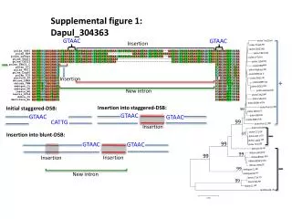
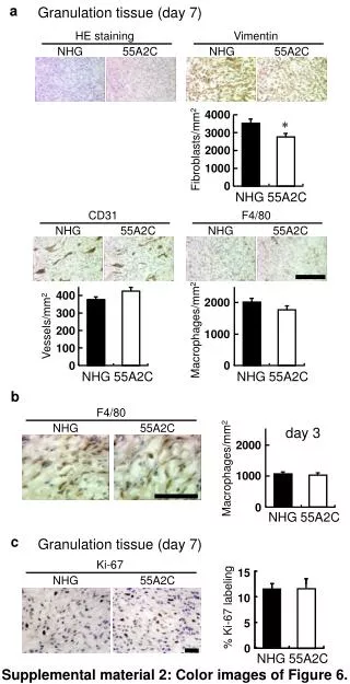
Explore the dynamic changes in granulation tissue on day 7 with HE staining, Vimentin, and macrophage/Fibroblast density analysis. Additionally, Ki-67 labeling percentage in fibroblasts. Supplementary material includes color images.

E N D
a Granulation tissue (day 7) HE staining Vimentin NHG 55A2C NHG 55A2C 4000 * 3000 Fibroblasts/mm2 2000 1000 0 NHG 55A2C CD31 F4/80 NHG 55A2C NHG 55A2C 400 2000 300 Vessels/mm2 Macrophages/mm2 200 1000 100 0 0 NHG 55A2C NHG 55A2C b F4/80 NHG 55A2C day 3 2000 Macrophages/mm2 1000 0 NHG 55A2C c Granulation tissue (day 7) Ki-67 15 NHG 55A2C 10 % Ki-67 labeling 5 0 NHG 55A2C Supplemental material 2: Color images of Figure 6.