Biochemistry
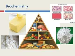
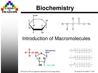
Biochemistry. Life’s Macromolecules. Life’s Macromolecules. Just as atoms can form bonds to make molecules, so too can molecules form bonds to make macromolecules Macromolecules are made by forming covalent bonds between repeating subunits called monomers .

Biochemistry
E N D
Presentation Transcript
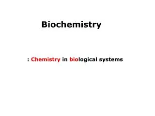
Biochemistry Life’s Macromolecules
Life’s Macromolecules • Just as atoms can form bonds to make molecules, so too can molecules form bonds to make macromolecules • Macromolecules are made by forming covalent bonds between repeating subunits called monomers. • Together these monomers make polymers or macromolecules • Carbohydrates • Proteins • Lipids • Nucleic acids
Making Macromolecules • A ‘building-up’ reaction is called an anabolic reaction, and requires the input of energy. • Condensation reactions (also called dehydration synthesis) occur which combines simple, low-energy molecules to more complex, high energy molecules • A byproduct of condensation reactions includes a molecule of water for each bond formed.
Breaking macromolecules • The reverse of a condensation reaction is a hydrolysis reaction (because water is required as a reactant) • These ‘breaking-down’ reactions are called catabolic reactions and usually releases energy
Carbohydrates • Monomers: monosaccharide : (single units of sugar) • e.g.: glucose, fructose, ribose, dihydroxyacetone
Monosaccharides • Can be classified by number of carbons, or by location of carbonyl group • In chain form, glucose has six carbons, and the carbonyl on a terminal carbon, so it is a hexose and an aldose. • Ribulose is a pentose and a ketose since it has five carbons and the carbonyl is on an internal carbon.
Monosaccharides • Dissolved in water, monosaccharides take on a ring shape • Glucose can form one of two isomers, depending on where the hydroxyl of carbon 1 ends up: above or below the plane of the ring
Disaccharides • Two monosaccharides attached with a covalent bond • e.g.: maltose, sucrose, lactose
Disaccharide formation • Consider two α–glucose molecules • This forms a 1-4 glycosidic bond
Polysaccharides • A chain of three or more sugars • Can form very long chains – used to store sugar molecules in cells • Animal cells: glycogen • Plant cells: amylose and amylopectin • Plant cells also form cellulose for the cell wall • Fungal cells use N-acetylglucosamine to form their cell wall material: chitin • Chitin is also found in the shells of lobsters, crabs and the exoskeleton of insects
Polysaccharide formation • Depending on which glucose molecules are used, different chain types can be created • Amylose uses α1-4 glycosidic bonds • Amylopectin and glycogen add branches to these chains with 1-6 glycosidic bonds
glycogen it’s big!
Cellulose • Uses 1-4 glycosidic linkages, which creates a straight chain, unlike the helical shape of amylose • Neighbouring chains can also form bonds, further strengthening the cellulose fibers. • These are then laid down in a woven fashion by the plant cell to create a very tough cell wall.
Lipids • Include fats, oils, waxes and sterols • Most common type of energy storage • Most common form in plants and animals is the triglyceride • All are hydrophobic • Store more than twice the energy per gram than carbohydrates or proteins
triglyceride • Also known as triacylglycerols
Oils vs fats • Saturated chain • Only single C-C bonds • Max number of H atoms • Overall straight chain shape • Higher melting/boiling pts. • Animal sources • Linked to heart disease • e.g.: butter, lard, shortening, hamburger fat • Unsaturated chain • At least 1 double C=C bond • Fewer H atoms • Overall bent chain shape • Lower melting/boiling pts. • Plant sources • Tend to be heart healthy • e.g.: olive oil, sunflower oil, peanut oil, palm oil FATS OILS
Waxes • Waxes are long hydrocarbon chains attached to either an alcohol (-OH) group or to rings of carbons • Are solid at room temperature but pliable • Useful for waterproof coatings • E.g.: • Waxy cuticle - on the surface of all land plants • Cerumen – the wax in human ears • Beeswax – used to make honeycomb in the beehive
Sterols • Several rings of carbon fused together • Used to make some hormones, as well as cholesterol cholesterol
Proteins • Monomer is the amino acid • Proteins are chains of amino acids which have folded over onto themselves to take on a unique shape • Shape is globular (rounded) or fibrous (stringy) • Enzymes, some hormones, membrane channels, and structural parts of cells are all made from protein • Aside from water, protein is the most abundant material in animals
Peptide Bond Formation • Proteins are long chains of amino acids • A condensation reaction occurs between the acid end of one amino acid and the amine end of another • This is repeated many times in reactions controlled by ribosomes • Ribosomes follow the genetic code to make a specific protein - the order and number of amino acids is unique to each protein, and is encoded in DNA
Structure of a polypeptide • Primary (1°) structure: the linear sequence of amino acids, which is unique to each • Secondary (2°) structure: even before the polypeptide chain has finished being created by the ribosomes, the amino acids begin to form intermolecular attractions which causes it to form either a helical shape (α-helix) or a folded shape (β-pleated sheet). • Tertiary (3°) structure: the R-groups interact with intermolecular forces to create a unique shape • Quaternary (4°) structure: some polypeptides will function with only 3° structure, but some must combine together with other polypeptides to become functional
A fibrous protein: collagen A globular protein: hemoglobin
Protein and shape • Shape is very important to how proteins function • For example, enzymes must be shaped correctly or they will not be able to combine with the molecule they break down. Enzyme (blue) must match shape of substrate (red) or chemical reaction cannot happen.
Protein and shape • Proteins are sensitive to changes in pH, as well as to increases in temperature • Both of these factors disrupt the intermolecular forces within a protein, causing them to change shape or denature • As long as the 1° structure is not lost, the polypeptide can return to it’s normal shape if the temperature or pH returns to normal • Coagulation occurs if the 1° structure is lost – this is a permanent change
Nucleic Acids • include molecules of heredity, energy carrier molecules, and some others (such as ribosomes) • made from monomers called nucleotides • Every nucleotide includes: • One sugar • One phosphate • One nitrogenous base
Energy carrier molecules ATP: adenosine triphosphate NADH: nicotinamide adenosine dinucleotide
Hereditary nucleic acids • DNA: deoxyribonucleic acid • one phosphate • deoxyribose sugar • one nitrogen base • Adenine • Guanine • Thymine • Cytosine • A pairs with T using two hydrogen bonds • G pairs with C using three hydrogen bonds Purines- double ringed Pyrimidines: single-ringed
Deoxyribonucleic acid • Backbone made of alternating sugars and phosphates, linked with phosphodiester bonds • complementary strands are anti-parallel • distance between bases when coiled into a double-helix is 0.34 nm • One complete turn of a strand is 3.4 nm • DNA turns in a right-handed direction (same as the threads on a wood screw)
Ribonucleic acid • mostly single-stranded • used in protein synthesis • mRNA – messenger RNA • rRNA – ribosomal RNA • tRNA – transfer RNA • Sugar is ribose, not deoxyribose • Nitrogen bases are the same except that the pyrimidine uracil is used instead of thymine