Download
1 / 15
160 likes | 376 Views
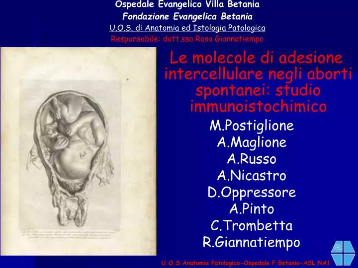
Ospedale Evangelico Villa Betania Fondazione Evangelica Betania U.O.S. di Anatomia ed Istologia Patologica Responsabile: dott.ssa Rosa Giannatiempo. U.O.S.Anatomia Patologica-Ospedale F.Betania-ASL NA1. Le molecole di adesione intercellulare negli aborti spontanei: studio immunoistochimico

E N D
Ospedale Evangelico Villa Betania Fondazione Evangelica Betania U.O.S. di Anatomia ed Istologia Patologica Responsabile: dott.ssa Rosa Giannatiempo U.O.S.Anatomia Patologica-Ospedale F.Betania-ASL NA1 Le molecole di adesione intercellulare negli aborti spontanei: studio immunoistochimico M.Postiglione A.Maglione A.Russo A.Nicastro D.Oppressore A.Pinto C.Trombetta R.Giannatiempo
U.O.S.Anatomia Patologica-Ospedale F.Betania-ASL NA1 Background Early placental development is associated with complex regulatory mechanisms, and molecular pathways involved during placental development are critical for pregnancy progression.
U.O.S.Anatomia Patologica-Ospedale F.Betania-ASL NA1 Objective We examined the role of adhesion molecules in the mechanism of spontaneous abortions.
U.O.S.Anatomia Patologica-Ospedale F.Betania-ASL NA1 STUDY DESIGN 2008:Materials II Trimestre 30 III Trimestre 20 I Trimestre 35 hhh
U.O.S.Anatomia Patologica-Ospedale F.Betania-ASL NA1 STUDY DESIGN 2008:Methods
U.O.S.Anatomia Patologica-Ospedale F.Betania-ASL NA1 CD31 U.O.S.Anatomia Patologica-Ospedale F.Betania-ASL NA1 Term placenta U.O.S.Anatomia Patologica-Ospedale F.Betania-ASL NA1 U.O.S.Anatomia Patologica-Ospedale F.Betania-ASL NA1
Results:CD31 Microvascular density (MVD) through CD31, decreased. Statistically significant differences have been detected between spontaneous abortion materials in the first trimester compared to those of abortions in the 2nd and the 3th trimesters with regard to cytotrophoblasts (CTs), syncytiotrophoblasts (STs), and extravillous trophoblasts (EVTs) (p<0.05). CD31-I Tr. U.O.S.Anatomia Patologica-Ospedale F.Betania-ASL NA1 CD31-I Tr. CD31-I Tr. U.O.S.Anatomia Patologica-Ospedale F.Betania-ASL NA1 U.O.S.Anatomia Patologica-Ospedale F.Betania-ASL NA1
CD31-II Tr. CD31-II Tr. U.O.S.Anatomia Patologica-Ospedale F.Betania-ASL NA1 U.O.S.Anatomia Patologica-Ospedale F.Betania-ASL NA1 CD31-III Tr. CD31-III Tr. U.O.S.Anatomia Patologica-Ospedale F.Betania-ASL NA1 U.O.S.Anatomia Patologica-Ospedale F.Betania-ASL NA1
CD44 U.O.S.Anatomia Patologica-Ospedale F.Betania-ASL NA1 Term placenta U.O.S.Anatomia Patologica-Ospedale F.Betania-ASL NA1
CD44-I Tr. Results:CD44 A down-regulation of CD44 was observed in decidual (D) cells. U.O.S.Anatomia Patologica-Ospedale F.Betania-ASL NA1 CD44-I Tr. CD44-I Tr. U.O.S.Anatomia Patologica-Ospedale F.Betania-ASL NA1 U.O.S.Anatomia Patologica-Ospedale F.Betania-ASL NA1
CD44-II Tr. CD44-II Tr. U.O.S.Anatomia Patologica-Ospedale F.Betania-ASL NA1 U.O.S.Anatomia Patologica-Ospedale F.Betania-ASL NA1 CD44-III Tr. CD44-III Tr. U.O.S.Anatomia Patologica-Ospedale F.Betania-ASL NA1 U.O.S.Anatomia Patologica-Ospedale F.Betania-ASL NA1
U.O.S.Anatomia Patologica-Ospedale F.Betania-ASL NA1 U.O.S.Anatomia Patologica-Ospedale F.Betania-ASL NA1 U.O.S.Anatomia Patologica-Ospedale F.Betania-ASL NA1 E-Cadherin Term placenta U.O.S.Anatomia Patologica-Ospedale F.Betania-ASL NA1
E-Cadherin-I Tr. Results:E-cadherin E-cadherin expression in cytotrophoblasts (CTs), and in syncytiotrophoblasts (STs),(p<0.05 and p<0.05), respectively, was significantly different. U.O.S.Anatomia Patologica-Ospedale F.Betania-ASL NA1 E-Cadherin-II Tr. E-Cadherin-III Tr. U.O.S.Anatomia Patologica-Ospedale F.Betania-ASL NA1 U.O.S.Anatomia Patologica-Ospedale F.Betania-ASL NA1
U.O.S.Anatomia Patologica-Ospedale F.Betania-ASL NA1 Decreased MVD in CTs in spontaneous abortion cases can suggest that MVD is critical in uteroplacental adequacy. Moreover,a down-regulation of E-cadherin could be responsible for impaired CTs differentiation and loss of the pregnancy. Furthermore, decreased CD44 expression in D cells may be cause of impairing stroma-villous connections. Their analysis in spontaneous abortions will provide useful information for clarifying the physiopathology of spontaneous abortions. Conclusions