Chapter 7 Protein synthesis
650 likes | 898 Views
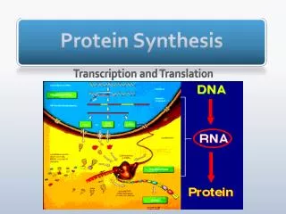
Chapter 7 Protein synthesis. 王心宇 副教授 College of Life Sciences. 8.1 Introduction. Figure 8.1 Size comparisons show that the ribosome is large enough to bind tRNAs and mRNA. 8.1 Introduction.

Chapter 7 Protein synthesis
E N D
Presentation Transcript
Chapter 7 Protein synthesis 王心宇 副教授 College of Life Sciences
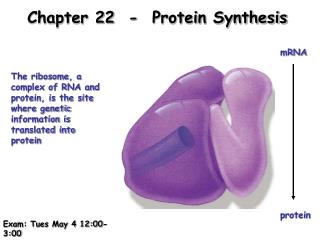
8.1 Introduction Figure 8.1Size comparisons show that the ribosome is large enough to bind tRNAs and mRNA.
8.1 Introduction Figure 8.2Ribosomes are large ribonucleoprotein particles that contain more RNA than protein and dissociate into large and small subunits.
8.2Protein synthesis occurs by initiation, elongation, and termination The A site of the ribosome is the site that an aminoacyl-tRNA enters to base pair with the codon. The P site of the ribosome is the site that is occupied by peptidyl-tRNA, the tRNA carrying the nascent polypeptide chain, still paired with the codon to which it bound in the A site. Figure 8.3The ribosome has two sites for binding charged tRNA.
8.2Protein synthesis occurs by initiation, elongation, and termination Figure 8.4The P and A sites position the two interacting tRNAs across both ribosome subunits.
8.2Protein synthesis occurs by initiation, elongation, and termination Figure 8.5 Aminoacyl-tRNA enters the A site, receives the polypeptide chain from peptidyl-tRNA, and is transferred into the P site for the next cycle of elongation.
8.2Protein synthesis occurs by initiation, elongation, and termination Figure 8.6tRNA and mRNA move through the ribosome in the same direction.
8.2Protein synthesis occurs by initiation, elongation, and termination Figure 8.7Protein synthesis falls into three stages.
8.2Protein synthesis occurs by initiation, elongation, and termination • Key concepts: • The ribosome has 3 tRNA-binding sites. • An aminoacyl-tRNA enters the A site. • Peptidyl-tRNA is bound in the P site. • Deacylated tRNA exits via the E site. • An amino acid is added to the polypeptide chain by transferring the polypeptide from peptidyl-tRNA in the P site to aminoacyl-tRNA in the A site.
8.3Special mechanisms control the accuracy of protein synthesis • Key concepts: • The accuracy of protein synthesis is controlled by specific mechanisms at each stage. Figure 8.8Errors occur at rates from 10-6 to 5 × 10⁻⁴ at different stages of protein synthesis
8.4Initiation in bacteria needs 30S subunits and accessory factors Initiation of protein synthesis is not a function of intact ribosomes, but is undertaken by the separate subunits, which reassociate during the initiation reaction. Figure 8.9Initiation requires free ribosome subunits. When ribosomes are released at termination, the 30S subunits bind initiation factors, and dissociate to generate free subunits. When subunits reassociate to give a functional ribosome at initiation, they release the factors.
8.4Initiation in bacteria needs 30S subunits and accessory factors • Initiation occurs at a special sequence on mRNA called the ribosome-binding site. The small and large subunits associate at the ribosome-binding site to form an intact ribosome. The reaction occurs in two steps: • Recognition of mRNA occurs when a small subunit binds to form an initiation complex at the ribosome-binding site. • Then a large subunit joins the complex to generate a complete ribosome.
8.4Initiation in bacteria needs 30S subunits and accessory factors Initiation factors (IF) (IF in prokaryotes, eIF in eukaryotes) are proteins that associate with the small subunit of the ribosome specifically at the stage of initiation of protein synthesis. Figure 8.10Initiation factors stabilize free 30S subunits and bind initiator tRNA to the 30S-mRNA complex.
8.4Initiation in bacteria needs 30S subunits and accessory factors • An initiation complex in bacterial protein synthesis contains a small ribosome subunit, initiation factors, and initiator aminoacyl-tRNA bound to mRNA at an AUG initiation codon. • IF-1 is a bacterial initiation factor that stabilizes the initiation complex. • IF-2 is a bacterial initiation factor that binds the initiator tRNA to the initiation complex. • IF-3 is a bacterial initiation factor required for 30S subunits to bind to initiation sites in mRNA. It also prevents 30S subunits from binding to 50S subunits.
8.4Initiation in bacteria needs 30S subunits and accessory factors Figure 8.11Initiation requires 30S subunits that carry IF-3.
8.4Initiation in bacteria needs 30S subunits and accessory factors • key concepts: • Initiation of protein synthesis requires separate 30S and 50S ribosome subunits. • Initiation factors (IF-1,2,3), which bind to 30S subunits, are also required. • A 30S subunit carrying initiation factors binds to an initiation site on mRNA to form an initiation complex. • IF-3 must be released to allow 50S subunits to join the 30S-mRNA complex.
8.5A special initiator tRNA starts the polypeptide chain • Synthesis of all proteins starts with the same amino acid: methionine. The signal for initiating a polypeptide chain is a special initiation codon that marks the start of the reading frame. Usually the initiation codon is the triplet AUG, but in bacteria, GUG or UUG are also used. • In bacteria and in eukaryotic organelles, the initiator tRNA carries a methionine residue that has been formylated on its amino group, forming a molecule of N-formyl-methionyl-tRNA. The tRNA is known as tRNAfMet. The name of the aminoacyl-tRNA is usually abbreviated to fMet-tRNAf.
8.5A special initiator tRNA starts the polypeptide chain N-formyl-methionyl-tRNA (tRNAfMet) is the aminoacyl-tRNA that initiates bacterial protein synthesis. The amino group of the methionine is formylated. tRNAmMet inserts methionine at internal AUG codons. Figure 8.12The initiator N-formyl-methionyl-tRNA (fMet-tRNAf) is generated by formylation of methionyl-tRNA, using formyl-tetrahydrofolate as cofactor.
8.5A special initiator tRNA starts the polypeptide chain Initiation in eukaryotes has the same general features as in bacteria. Initiation in eukaryotic cytoplasm uses AUG as the initiator. The initiator tRNA is a distinct species, but its methionine does not become formylated. It is called tRNAiMet. So the difference between the initiating and elongating Met-tRNAs lies solely in the tRNA moiety, with Met-tRNAi used for initiation and Met-tRNAm used for elongation. The two tRNAs are distinguished by their tertiary structures and also by a phosphorylation of the 2 ribose position on base 64 of the initiator.
8.6Use of fMet-tRNAf is controlled by IF-2 and the ribosome The initiation reaction involves binding of a 30S subunit to a ribosome-binding site. The two features of a bacterial ribosome-binding site are the AUG initiation codon and a polypurine sequence preceding it by ~10 bases that corresponds to the hexamer 5’ ... A G G A G G... 3’. This polypurine stretch is known as the Shine-Dalgarno sequence. It is complementary to a highly conserved sequence close to the 3’ end of 16S rRNA. Written in reverse direction, the rRNA sequence is the hexamer: 3’ ... U C C U C C... 5’
8.6Use of fMet-tRNAf is controlled by IF-2 and the ribosome Figure 8.13Only fMet-tRNAf can be used for initiation by 30S subunits; only other aminoacyl-tRNAs (αα-tRNA) can be used for elongation by 70S ribosomes.
8.6Use of fMet-tRNAf is controlled by IF-2 and the ribosome Figure 8.14IF-2 is needed to bind fMet-tRNAf to the 30S-mRNA complex. After 50S binding, all IF factors are released and GTP is cleaved.
8.6Use of fMet-tRNAf is controlled by IF-2 and the ribosome • key concepts • An initiation site on bacterial mRNA consists of the AUG initiation codon preceded with a gap of ~10 bases by the Shine-Dalgarno polypurine hexamer. • The rRNA of the 30S bacterial ribosomal subunit has a complementary sequence that base pairs with the Shine-Dalgarno sequence during initiation. • IF-2 binds the initiator fMet-tRNAf and allows it to enter the partial P site on the 30S subunit.
8.7Small subunits scan for initiation sites on eukaryotic mRNA Initiation of protein synthesis in eukaryotic cytoplasm resembles the process in bacteria, but the order of events is different, and the number of accessory factors is greater. Figure 8.15Eukaryotic ribosomes migrate from the 5’ end of mRNA to the ribosome binding site, which includes an AUG initiation codon.
8.7Small subunits scan for initiation sites on eukaryotic mRNA Eukaryotic initiation factors are named similarly to those in bacteria, sometimes by analogy with the bacterial factors, and are given the prefix "e" to indicate their eukaryotic origin. They act at all stages of the process, including: • forming an initiation complex with the 5’ end of mRNA • forming a complex with Met-tRNAi • binding the mRNA-factor complex to the Met-tRNAi-factor complex • enabling the ribosome to scan mRNA from the 5’ end to the first AUG • detecting binding of initiator tRNA to AUG at the start site • mediating joining of the 60S subunit.
8.7Small subunits scan for initiation sites on eukaryotic mRNA Figure 8.16Some initiation factors bind to the 40S ribosome subunit to form the 43S complex; others bind to mRNA. When the 43S complex binds to mRNA, it scans for the initiation codon and can be isolated as the 48S complex.
8.7Small subunits scan for initiation sites on eukaryotic mRNA • key concepts • Eukaryotic 40S ribosomal subunits bind to the 5’ end of mRNA and scan the mRNA until they reach an initiation site. • A eukaryotic initiation site consists of a 10 nucleotide sequence that includes an AUG codon. • Initiation factors are required for all stages of initiation, including binding the initiator tRNA, 40S subunit attachment to mRNA, movement along the mRNA, and joining of the 60S subunit. • eIF2 and eIF3 bind the initiator Met-tRNAi and GTP, and the complex binds to the 40S subunit before it associates with mRNA. • 60S ribosomal subunits join the complex at the initiation site.
8.8Elongation factor Tu loads aminoacyl-tRNA into the A site Once the complete ribosome has formed at the initiation codon, with initiator Met-tRNA in the P site, the A site is ready to accept an aminoacyl-tRNA. Any aminoacyl-tRNA except the initiator can enter the A site. Its entry is mediated by an elongation factor (EF-Tu in bacteria). The process is similar in eukaryotes. Figure 8.17EF-Tu-GTP places aminoacyl-tRNA on the ribosome and then is released as EF-Tu-GDP. EF-Ts is required to mediate the replacement of GDP by GTP. The reaction consumes GTP and releases GDP. The only aminoacyl-tRNA that cannot be recognized by EF-Tu-GTP is fMet-tRNAf, whose failure to bind prevents it from responding to internal AUG or GUG codons.
8.8Elongation factor Tu loads aminoacyl-tRNA into the A site • Key Terms • Elongation factors (EF in prokaryotes, eEF in eukaryotes) are proteins that associate with ribosomes cyclically, during addition of each amino acid to the polypeptide chain. • EF-Tu is the elongation factor that binds aminoacyl-tRNA and places it into the A site of a bacterial ribosome. • Key Concepts • EF-Tu·GTP binds aminoacyl-tRNA and places it in the ribosome A site. • GTP is hydrolysis is needed to release EF-Tu after the aminoacyl-tRNA has paired with its codon.
8.9The polypeptide chain is transferred to aminoacyl-tRNA Peptidyl transferase is the activity of the ribosomal 50S subunit that synthesizes a peptide bond when an amino acid is added to a growing polypeptide chain. The actual catalytic activity is a property of the rRNA. Figure 8.18Peptide bond formation takes place by reaction between the polypeptide of peptidyl-tRNA in the P site and the amino acid of aminoacyl-tRNA in the A site.
8.9The polypeptide chain is transferred to aminoacyl-tRNA Puromycin is an antibiotic that terminates protein synthesis by mimicking a tRNA and becoming linked to the nascent protein chain. Figure 8.19Puromycin mimics aminoacyl-tRNA because it resembles an aromatic amino acid linked to a sugar-base moiety.
8.9The polypeptide chain is transferred to aminoacyl-tRNA • Key Concepts • The peptidyl transferase activity of the 50S subunit transfers the nascent polypeptide chain from peptidyl-tRNA in the P site to aminoacyl-tRNA in the A site. • Peptide bond synthesis generates deacylated tRNA in the P site and peptidyl-tRNA in the A site.
8.10Translocation moves the ribosome Translocation is the movement of the ribosome one codon along mRNA after the addition of each amino acid to the polypeptide chain. Figure 8.20A bacterial ribosome has 3 tRNA-binding sites. Aminoacyl-tRNA enters the A site of a ribosome that has peptidyl-tRNA in the P site. Peptide bond synthesis deacylates the P site tRNA and generates peptidyl-tRNA in the A site. Translocation moves the deacylated tRNA into the E site and moves peptidyl-tRNA into the P site.
8.10Translocation moves the ribosome Most thinking about translocation follows the hybrid state model, which proposes that translocation occurs in two stages. Figure 8.21Models for translocation involve two stages. First, at peptide bond formation the aminoacyl end of the tRNA in the A site becomes located in the P site. Second, the anticodon end of the tRNA becomes located in the P site.
8.10Translocation moves the ribosome • key concepts • Ribosomal translocation moves the mRNA through the ribosome by 3 bases, moving deacylated tRNA into the E site, moving peptidyl-tRNA into the P site, and emptying the A site. • The hybrid state model proposes that translocation occurs in two stages, in which the 50S moves relative to the 30S, and then the 30S moves along mRNA to restore the original conformation.
8.11Elongation factors bind alternately to the ribosome Translocation requires GTP and another elongation factor, EF-G. This factor is a major constituent of the cell; it is present at a level of ~1 copy per ribosome (~20,000 molecules per cell).EF-G is an elongation factor needed for the translocation stage of bacterial protein synthesis. Figure 8.22Binding of factors EF-Tu and EF-G alternates as ribosomes accept new aminoacyl-tRNA, form peptide bonds, and translocate.
8.11Elongation factors bind alternately to the ribosome • EF-G requires GTP to function in translocation, and has a structure resembling the aminoacyl-tRNA·EF-Tu·GTP complex. • Binding of EF-Tu and EF-G to the ribosome is mutually exclusive. • Translocation requires GTP hydrolysis, which triggers a change in EF-G, which in turn triggers a change in ribosome structure. Figure 8.23The structure of the ternary complex of aminoacyl-tRNA·EF-Tu·GTP (left) resembles the structure of EF-G (right). Structurally conserved domains of EF-Tu and EF-G are in red and green; the tRNA and the domain resembling it in EF-G are in purple.
8.12Uncharged tRNA causes the ribosome to trigger the stringent response The ribosome is not merely the key complex that synthesizes proteins, but also triggers several types of regulatory response. The initial stimulus for these responses is the absence of amino acids (resulting from poor growth conditions), which in turn leads to a depletion of aminoacyl-tRNA. The ribosome is in a key position to detect a deficiency in aminoacyl-tRNA, because this stops it from functioning in protein synthesis. Lack of aminoacyl-tRNAs in general is used to trigger an alarm response; also, the absence of a specific aminoacyl-tRNA can be used to regulate the metabolic systems that produce the corresponding amino acid.
8.12Uncharged tRNA causes the ribosome to trigger the stringent response When bacteria find themselves in such poor growth conditions that they lack a sufficient supply of amino acids to sustain protein synthesis, they shut down a wide range of activities. This is called the stringent response. The stringent response is controlled by the accumulation of two unusual nucleotides. ppGpp is guanosine tetraphosphate, with diphosphates attached to both 5’ and 3’ positions. pppGpp is guanosine pentaphosphate, with a 5 triphosphate group and a 3 diphosphate. These nucleotides are typical small-molecule effectors that function by binding to target proteins to alter their activities. Sometimes they are known collectively as (p)ppGpp.
8.12Uncharged tRNA causes the ribosome to trigger the stringent response Bacterial mutants that cannot produce the stringent reponse are called relaxed. The most common site of relaxed mutation lies in the gene relA, which codes for a protein called the stringent factor. The presence of uncharged tRNA in the A site blocks protein synthesis and it triggers an idling reaction by wild-type ribosomes. Provided that the A site is occupied by an uncharged tRNA specifically responding to the codon, the RelA protein catalyzes a reaction in which ATP is used to donate a pyrophosphate group to the 3’ position of either GTP or GDP. The formal name for this activity is (p)ppGpp synthetase.
8.12Uncharged tRNA causes the ribosome to trigger the stringent response Figure 8.24Stringent factor catalyzes the synthesis of pppGpp and ppGpp; ribosomal proteins can dephosphorylate pppGpp to ppGpp.
8.12Uncharged tRNA causes the ribosome to trigger the stringent response The presence of uncharged tRNA in the A site blocks protein synthesis and it triggers an idling reaction by wild-type ribosomes. Figure 8.25In normal protein synthesis, the presence of aminoacyl-tRNA in the A site is a signal for peptidyl transferase to transfer the polypeptide chain, followed by movement catalyzed by EF-G; but under stringent conditions, the presence of uncharged tRNA causes RelA protein to synthesize (p)ppGpp and to expel the tRNA.
8.12Uncharged tRNA causes the ribosome to trigger the stringent response • ppGpp is an effector for controlling several reactions, including the inhibition of transcription. In particular, it specifically inhibits transcription at the promoters of operons coding for rRNA. More generally, it causes the rate of transcription to decrease. Together these effects account for the ability of the stringent response to greatly reduce the energy that the cell spends on gene expression. • key concepts • Poor growth conditions cause bacteria to produce the small molecule regulators ppGpp and pppGpp. • The trigger for the reaction is the entry of uncharged tRNA into the ribosomal A site, which activates the (p)ppGpp synthetase of the stringent factor RelA. • One (p)ppGpp is produced every time an uncharged tRNA enters the A site.
8.13Three codons terminate protein synthesis and are recognized by protein factors A stop codon(Termination codon) is one of three triplets (UAG, UAA, UGA) that causes protein synthesis to terminate. They are also known historically as nonsense codons. Two stages are involved in ending translation. The termination reaction itself involves release of the protein chain from the last tRNA. The post-termination reaction involves release of the tRNA and mRNA, and dissociation of the ribosome into its subunits. None of the termination codons is represented by a tRNA. They function in an entirely different manner from other codons, and are recognized directly by protein factors. (Since the reaction does not depend on codon-anticodon recognition, there seems to be no particular reason why it should require a triplet sequence. Presumably this reflects the evolution of the genetic code.)
8.13Three codons terminate protein synthesis and are recognized by protein factors • A release factor (RF) is required to terminate protein synthesis to cause release of the completed polypeptide chain and the ribosome from mRNA. Individual factors are numbered. Eukaryotic factors are called eRF. • RF1 is the bacterial release factor that recognizes UAA and UAG as signals to terminate protein synthesis. • RF2 is the bacterial release factor that recognizes UAA and UGA as signals to terminate protein synthesis. • RF3 is a protein synthesis termination factor related to the elongation factor EF-G. It functions to release the factors RF1 or RF2 from the ribosome when they act to terminate protein synthesis.
8.13Three codons terminate protein synthesis and are recognized by protein factors The factors act at the ribosomal A site and require polypeptidyl-tRNA in the P site. They activate the ribosome to hydrolyze the peptidyl tRNA. Cleavage of polypeptide from tRNA takes place by a reaction analogous to the usual peptidyl transfer, except that the acceptor is H2O instead of aminoacyl-tRNA. Figure 8.26Molecular mimicry enables the elongation factor Tu-tRNA complex, the translocation factor EF-G, and the release factors RF1/2-RF3 to bind to the same ribosomal site.
8.13Three codons terminate protein synthesis and are recognized by protein factors The eukaryotic class 1 release factor, eRF1, is a single protein that recognizes all three termination codons. Figure 8.27The eukaryotic termination factor eRF1 has a structure that mimics tRNA. The motif GGQ at the tip of domain 2 is essential for hydrolyzing the polypeptide chain from tRNA.
8.13Three codons terminate protein synthesis and are recognized by protein factors dissociation of the remaining components (tRNA, mRNA, 30S and 50S subunits) requires the factor RRF, ribosome recycling factor. This acts together with EF-G in a reaction that uses hydrolysis of GTP. Figure 8.28The RF (release factor) terminates protein synthesis by releasing the protein chain. The RRF (ribosome recycling factor) releases the last tRNA, and EF-G releases RRF, causing the ribosome to dissociate.
8.13Three codons terminate protein synthesis and are recognized by protein factors • key concepts • The three termination codons are used in bacteria with relative frequencies UAA>UGA>UAG. • Termination codons are recognized by protein release factors, not by aminoacyl-tRNAs. • The structures of the class 1 release factors (RF1 and RF2 in E. coli) resemble aminoacyl-tRNA·EF-Tu and EF-G. • The class 1 release factors respond to specific termination codons and hydrolyze the polypeptide-tRNA linkage. • The class 1 release factors are assisted by class 2 release factors (such as RF3) that depend on GTP. • The mechanism is similar in bacteria (which have two types of class 1 release factors) and eukaryotes (which have only one class 1 release factor).
8.14Ribosomal RNA pervades both ribosomal subunits • key concepts • Each rRNA has several distinct domains that fold independently. • Virtually all ribosomal proteins are in contact with rRNA. • Most of the contacts between ribosomal subunits are made between the 16S and 23S rRNAs.