Download
1 / 79
870 likes | 1.51k Views
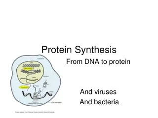
Chapter 30 Protein Synthesis. Outline. The genetic code. Matching an amino acid with its proper tRNA. Condon-anticodon pairing. Structure and assembly of ribosomes. Events in mRNA translation. Proteins synthesis in eukaryotic cells. A Generalized Secondary Structure of tRNA Molecules.

E N D
Outline • The genetic code. • Matching an amino acid with its proper tRNA. • Condon-anticodon pairing. • Structure and assembly of ribosomes. • Events in mRNA translation. • Proteins synthesis in eukaryotic cells.
A Generalized Secondary Structure of tRNA Molecules Figure 30.1 Generalized secondary structure of tRNA molecules. Circles represent nucleotides in the tRNA sequence. The numbers given indicate the standardized numbering for RNAs. Dots indicated places where the number of nucleotides may vary in different tRNA species.
30.1 The Genetic Code • The genetic code is a triplet code, read from a fixed starting point in each mRNA. • A group of 3 bases (a codon) codes for one amino acid. • The code is not overlapping. • All the codons have meaning. • The genetic code is degenerate – in most cases, each amino acid can be coded for by any of several triplet codons. • The base sequence is read from a fixed starting point without punctuation. • The genetic code is “universal”.
Features of the Code Figure 30.2 (a) An overlapping versus a nonoverlapping code. (b) A continuous versus a punctuated code.
30.2 The Genetic Code *Translation initiator as well as Met.
30.2 Matching an Amino Acid with Its Proper tRNA • Codon recognition is achieved by aminoacyl-tRNAs. • For accurate translation to occur, the appropriate aminoacyl-tRNA must “read” the codon through base pairing via its anticodon loop. • The code by which each aminoacyl-tRNA synthetase matches up its amino acid with tRNAs constitutes a second genetic code. • Aminoacyl-tRNA synthetases interpret the second genetic code. • Aminoacyl-tRNA synthetases discriminate between the various tRNAs and amino acids.
Evolution Has Provided Two Distinct Classes of Aminoacyl-tRNA Synthetases • Aminoacyl-tRNA synthetases fall into two fundamental classes on the basis of similar amino acid sequence motifs, oligomeric state, and acylation function. • Class I aminoacyl-tRNA synthetases first add the amino acid to the 2'-OH of the terminal adenylate residue of tRNA before shifting it to the 3'-OH. • Class II enzymes add it directly to the 3'-OH (see Figure 30.3).
The Aminoacyl-tRNA Synthetase Reaction Figure 30.3 (a) the overall reaction. (b) Aminoacyl-tRNA formation proceeds in two steps: (i) formation of the aminoacyl-adenylate and (ii) transfer of the activated amino acid to either the 2'-OH or 3'-OH.
The Aminoacyl-tRNA Synthetase Reaction Figure 30.3
tRNA Molecules Have an Acceptor Stem at One End and an Anticodon at the Other Figure 30.5 Ribbon diagram of the tRNA tertiary structure.
tRNA Recognition For most tRNAs, a set of sequence elements is recognized by its specific aminoacyl-tRNA synthtase, rather than a single distinctive nucleotide or base pair. Figure 30.6 Major identity elements in four tRNA species. Numbered filled circles indicate positions of identity elements within the tRNA that are recognized by its specific aminoacyl-tRNA synthetase.
Structure of an E. coli Glutaminyl-tRNA Synthetase Complexed with tRNA Figure 30.7 Structure of E. coli glutaminyl-tRNAGln synthetase complexed with tRNAGln and ATP. The protein: tRNA contact region extends along one side of the entire length of this extended protein. The acceptor stem of the tRNA and the ATP (green) fit into a cleft at the top of the protein in this view. The enzyme also interacts extensively with the anticodon (lower tip of tRNAGln).
A Single G:U Base Pair Defines tRNAAlas Figure 30.8 (a) a microhelix analog of tRNAAla is aminoacylated by alanyl-tRNAAla synthetase, provided it has the characteristic tRNAAla G3:U70 acceptor stem base pair.
30.3 Codon-Anticodon Pairing Figure 30.9 Codon-anticodon pairing. Complementary trinucleotide sequence elements align in antiparallel fashion.
30.3 Codon-Anticodon Pairing • Francis Crick proposed the “wobble” hypothesis for codon:anticodon pairing. • He hypothesized that the first two bases of the codon and the last two bases of the anticodon form canonical Watson-Crick base pairs. • But pairing between the third base of the codon and the first base of the anticodon follows less stringent rules. • That is, there is play or wobble in base pairing at this position.
30.3 Codon-Anticodon Pairing Figure 30.10 Pairing of anticodon inosine (I is on the left) with C, U, or A as the codon third base. Note that I is the keto tautomeric form.
Some Codons Are Used More Than Others • Because more than one codon exists for most amino acids, variation in codon usage is possible. • Thus the DNA of different organisms varies in relative A:T/G:C content. • Even in organisms of average base composition, codon usage may be biased. • Examples of nonrandom usage of codons in E. coli and in humans are given in Table 30.4. • Preferred codons are represented by the most abundant isoacceptor tRNAs. • And mRNAs for proteins that are synthesized in abundance tend to employ preferred codons.
Nonsense Suppression Occurs When Suppressor tRNAs Read Nonsense Codons • Mutations that alter a sense codon to one of the three nonsense (stop) codons, UAA (ochre), UAG (amber), or UGA (opal), can result in premature termination of protein synthesis and the release of truncated (incomplete) proteins. • It has been found that second mutations elsewhere in the genome can suppress the effects of nonsense mutations, so that the organism survives. This phenomenon is termed nonsense suppression. • Suppressors are mutations in tRNA genes that change the anticodon so that the mutant tRNA is now able read a particular “stop” codon and insert an amino acid.
30.4 Structure and Assembly of Ribosomes • Protein biosynthesis occurs by the process of translation on ribosomes. • Ribosomes are compact ribonucleoprotein particles found in the cytosol of all cells. • The E. coli ribosome is 25 nm diameter, 2520 kD in mass, and consists of two unequal subunits that dissociate at < 1mM Mg2+. • 30S subunit is 930 kD with 21 proteins and a 16S rRNA. • 50S subunit is 1590 kD with 31 proteins and two rRNAs: 23S rRNA and 5S rRNA. • The 30S and 50S subunits combine to form the 70S ribosome.
30.4 Structure and Assembly of Ribosomes * S proteins. †L proteins. Ribosomes are roughly 2/3 RNA. There are approximately 20,000 ribosomes in a cell representing 20% of cell's mass.
The rRNAs of E. coli Are Encoded by a Set of Seven Operons Example: Pre-rRNA from one of the seven rRNA operons. Figure 30.11 E. coli has seven ribosomal RNA operons. Numerals to the right of the brackets indicate the number of species of tRNA encoded by each transcript.
Prokaryotic Ribosomes Are Made from 50 Different Proteins and Three Different RNAs • There is one copy of each ribosomal protein per 70S ribosome, except L7/L12 with 4. (L = large subunit) • Proteins L7/L12 are identical except for extent of acetylation at N-terminus. • Four L7/L12 plus L10 makes "L8". • Only one protein is common to large and small subunits: S20 = L26. (S = small subunit) • The largest ribosomal protein is S1 (557 residues, 61.2 kD). • The smallest ribosomal protein is L34 (46 residues, 5.4 kD).
Ribosomes Self-Assemble Spontaneously in Vitro • Ribosome subunit self-assembly is one of the paradigms for the spontaneous formation of supramolecular complexes. • Structures of large and small subunits are known. • If individual proteins and rRNAs are mixed, functional ribosomes will assemble. • rRNA acts as a scaffold on which the ribosomal proteins convene in a specific order. • A tunnel runs through the large subunit . • Growing peptide chain is threaded through the tunnel during protein synthesis.
The Overall Form of the 30S Structure is Essentially That of the rRNA Figure 30.13 Structure of T. thermophilus ribosomal subunits and 70S ribosome. Features are labeled. (a) 30S; (b) 50S; (c) 70S; (d) side view of 70S.
Ribosomes Have a Characteristic Anatomy • The details of ribosome structure have been revealed in recent years. • The 30S subunit features a “head” and a “body”, from which a “platform” projects. • The head, body, and platform create a cleft between them. • The 50S subunit is a mitt-like globular structure with three distinct projections: a “central protuberance”, the “stalk”, and a ridge known as the “L7/L12 ridge”. • The catalytic center on 50S, the peptidyl transferase, is located at the bottom of a deep cleft in 50S. • Both subunits are involved in translocation, in which mRNA moves through the ribosome.
The Cytosolic Ribosomes of Eukaryotes Are Larger than Prokaryotic Ribosomes • Mitochondrial and chloroplast ribosomes are quite similar to prokaryotic ribosomes, reflecting their prokaryotic origin. • Eucaryotic cytoplasmic ribosomes are larger and more complex than procaryotic, but many of the structural and functional properties are similar. • The structural organization of mammalian ribosomes is described in Table 30.6.
The Cytosolic Ribosomes of Eukaryotes Are Larger than Prokaryotic Ribosomes
30.5 Events in mRNA Translation • All protein synthesis involves three phases: initiation, elongation, and termination. • Initiation involves binding of mRNA and initiator aminoacyl-tRNA to small subunit, followed by binding of large subunit. • Elongation: Movement of ribosome along mRNA and synthesis of all peptide bonds - with tRNAs bound to acceptor (A) and peptidyl (P) sites. See Figure 30.14. • Termination occurs when stop codon is reached. • Three tRNA molecules may be associated with the ribosome:mRNA complex at any moment.
30.5 Events in mRNA Translation Figure 30.14 The basic steps in protein synthesis. The ribosome has three distinct binding sites for tRNA: the A, or acceptor, site; the P, or peptidyl, site; and the E, or exit, site.
30.5 Events in mRNA Translation Figure 30.14 The basic steps in protein synthesis. The ribosome has three distinct binding sites for tRNA: the A, or acceptor, site; the P, or peptidyl, site; and the E, or exit, site.
30.5 Events in mRNA Translation Figure 30.14 The basic steps in protein synthesis. The ribosome has three distinct binding sites for tRNA: the A, or acceptor, site; the P, or peptidyl, site; and the E, or exit, site.
30.5 Events in mRNA Translation Figure 30.15 The three tRNA binding sites on ribosomes. The view shows the ribosomal surfaces that form the interface between the (a) 30S and (b) 50S subunits in a 70S ribosome. The A (green), P (blue), and E (yellow) sites have bound tRNA.
Peptide Chain Initiation in Prokaryotes • The components required for peptide chain initiation include (1) mRNA; (2) 30S and 50S ribosomal subunits; (3) a set of proteins known as initiation factors; (4) GTP; and f-Met-tRNAifMet. • The initiator tRNA is f-Met-tRNAifMet, has formylMet. • A formyl transferase adds the formyl group (see Figure 30.17). • N-formyl-Met-tRNAifMet is only used for initiation, and regular Met-tRNAmfMet is used for incorporation of Met internally. • N-formyl methionine is first amino acid of all E.coli proteins and the N-terminal Met is removed post-translationally in many proteins.
Initiator tRNA Figure 30.16 The secondary structure of E. coli N-formyl-methionyl-tRNAifMet. The features distinguishing it from non-initiator tRNAs are highlighted.
The Transformylation of Methionyl-tRNAifMet Figure 30.17 Methionyl-tRNAifMet formyl transferase catalyzes the transformylation of methionyl-tRNAifMet using N10-formyl-THF as formyl donor. The tRNA for reading Met codons within a protein (tRNAMet) is not a substrate for this transformylase. Eucaryotes do not use formyl-Met-tRNA.
mRNA Recognition and Alignment • Correct positioning of mRNA on ribosome requires alignment of a pyrimidine-rich sequence on 3'-end of 16S RNA with a purine-rich part of 5'-end of mRNA. • The purine-rich segment - the ribosome-binding site - is known as the Shine-Dalgarno sequence, AGGA (see Figure 30.18). (No SD sequence in eucaryotes.) • Initiation factor proteins, GTP, N-formyl-Met- tRNAifMet, mRNA and 30S ribosome combine to form the 30S initiation complex. • The 50S subunit then binds to form a 70S initiation complex. • The initiation factors are soluble proteins required for assembly of these initiation complexes (Table 30.7).
Various Shine-Dalgarno Sequences Recognized by E. coli Ribosomes Figure 30.18 These sequences lie about ten nucleotides upstream from their respective AUG initiation codon and are complementary to the UCCU core sequence element of E. coli 16S rRNA.
Properties of E. coli Initiation Factors Table 30.8 Protein initiation factors. IF-2 ia a G-protein as are EF-Tu, EF-G and RF 3.
Events of Initiation • 30S subunit with IF-1 and IF-3 binds mRNA, IF-2, GTP and f-Met-tRNAifMet (Figure 30.19). • IF-2 delivers the initiator tRNA in a GTP-dependent process. • Loss of the initiation factors leads to binding of 50S subunit. • Note that the "acceptor site" is now poised to accept an incoming aminoacyl-tRNA.
The Sequence of Events in Peptide Chain Initiation Figure 30.19 Initiation begins when a 30S subunit:(IF-3:IF-1) complex binds mRNA and a complex of IF-2, GTP, and f-Met-tRNAifMet.
Peptide Chain Elongation Requires Two G-Protein Family Members The requirements for peptide chain elongation are: • An mRNA:70S ribosome:peptidyl-tRNA complex (peptidyl-tRNA in the P site). • Aminoacyl-tRNAs. • A set of proteins known as elongation factors. • GTP.
Peptide Chain Elongation Requires Two G-Protein Family Members Chain elongation can be divided into 3 steps: • Codon-directed binding of the incoming aminoacyl-tRNA at the A site. Decoding center regions of 16S rRNA make sure the proper aminoacyl-tRNA is in the A site. • Peptide bond formation: transfer of the peptidyl chain from the tRNA bearing it to the –NH2 group of the new amino acid. • Translocation of the “one-residue-longer” peptidyl-tRNA to the P site to make room for the next aminoacyl-tRNA at the A site. These shifts are coupled with movement of the ribosome one codon further along the mRNA.
Peptide Chain Elongation Requires Two G-Protein Family Members • Protein synthesis is vital to cell function, so elongation factors are present in significant quantities (EF-Tu is 5% of total protein in E. coli). • EF-Tu binds aminoacyl-tRNA and GTP. • Aminoacyl-tRNA binds to A site of ribosome as a complex with 2EF-Tu and 2GTP. • GTP is then hydrolyzed and the EF-Tu:GDP complex dissociate. • EF-Ts is a guanine nucleotide exchange factor (GEF) and it recycles EF-Tu by exchanging GTP for GDP.
Peptide Chain Elongation Requires Two G-Protein Family Members Table 30.8 Protein elongation factors.
Peptidyl Transfer – the Central Reaction of Protein Synthesis • Peptidyl transfer, or transpeptidation, is the actual peptide bond-forming step. • No energy input is needed here; the ester linking the peptidyl moiety to tRNA is intrinsically reactive. • Figure 30.21 shows the location of the decoding center and highlights the interface between a codon and anticodon. • The peptidyl transferase, the activity catalyzing peptide bond formation, is associated with the 50S ribosomal subunit. • The reaction is part of the 23S rRNA in the 50S.
Figure 30.20 The cycle of events in peptide chain elongation of E. coli ribosomes. The structure at lower left is an EF-Tu:aminoacyl-tRNA complex.