
Nervous system
Nervous system. Nervous system - Outline. Nervous tissue Central Nervous System = CNS Brain Spinal cord Peripheral Nervous System = PNS Cranial nerves Spinal nerves Autonomic Nervous System (a special case). The spinal cord. Division based on the vertebrae: cervical (C1-C7)

Nervous system
E N D
Presentation Transcript
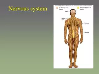
Nervous system - Outline • Nervous tissue • Central Nervous System = CNS • Brain • Spinal cord • Peripheral Nervous System = PNS • Cranial nerves • Spinal nerves • Autonomic Nervous System (a special case)
The spinal cord • Division based on the vertebrae: • cervical (C1-C7) • Thoracic (T1-T12) • lumbar (L1-L5) • sacral (S1-S5= sacrum) • coccygeal (Co1-Co4=coccyx) • The lower end of the spinal cord is pulled up during growth, but the nerves exit through the same foramen formation of the cauda equina
The spinal chord • Anatomy: • Cylinder of nervous tissue • From the foramen magnum to lumbar vertebae 1 (L1) • During growth, vertebrae grow faster than nervous tissue spinal cord “moves up” the vertebrae • The meningeal sac continues down the vertebral canal where it is filled only with CSF ideal place for lumbar puncture
The three spinal meningeal layers • Dura mater: Most superficial layer • Arachnoid mater: just inside the dura • Simple squamous epithelial cells • Between these and the dura mater is the subarachnoid space that is filled with cerebrospinal fluid • Pia mater: surrounding the spinal chord proper • Very delicate and clear • Is anchored to the arachnoid mater by small processes
Spinal meninges • Unlike cranial meninges, there is a space between the dura mater and the vertebra epidural space where epidural injections are done • The anesthetic injection remains around the nerve root anesthesia at that level • Intrathecal injections are given within the subarachnoid space
The spinal cord • Spinal cord has two parts: • White matter (axons and dendrites (white matter due to the myelin sheaths): Conduct signals to and from the brain • Grey matter: Reflexes (due to the neuron body and unmyelinated sheaths) • So what is a reflex? A set of preprogrammed reactions responding to a specific signal
Grey matter of the spinal cord • Located toward the center and surrounded by white matter • Dorsal horn: interneurons • Lateral horn: first motor neurons of the ANS • Ventral horn: motor neurons of the somatic NS (for skeletal muscles), also know as the lower motor neurons
White matter of the spinal cord • Axons and dendrites arriving and leaving the spinal cord • Arranged in bundles of fibers with common purpose • Sensory fibers (from body to brain) arrive in the spinal cord through the posterior root • The sensory neuron is itself located in the dorsal root ganglion • The motor fibers (from spinal cord to body) are leaving through the ventral roots. • Note that both ventral and dorsal roots join just before entering the transverse foramina and form the spinal nerves
White matter of the spinal cord • The ascending pathways (sensory bundles) are found in the posterior aspect and lateral aspects of the spinal cord • The descending pathways (motor bundles) are found laterally/internally and ventrally in the spinal cord
Spinal “columns/tracts” • Most of the tracts traveling through the spinal cord decussate cross-over to the other side • Parts of the left side of the brain control the right side of your body etc. • “Contralateral” = origin of the signal (be it sensory or motor) is on the opposite side of the target • Right side of brain controlling left side of body • “Ipsilateral” = origin of the signal is on the SAME side of the body • Right side of brain controlling right side of body
Shingles / Chicken pox • Shingles (Herpes zoster) is a virus that lies dormant in the dorsal root ganglion for life • If you’ve had chicken pox, you have this virus in your spinal chord!!!!! • If left unchecked by your immune system, this virus will travel back down the nerves and erupt in the corresponding area shingles • Swelling on 1 side, near spine (dorsal root ganglia swell with fluid) • Treatment: You must go on antiviral quickly to minimize the disease. Otherwise, the course is very long and very painful
Nervous system - Outline • Nervous tissue • Central Nervous System = CNS • Brain • Spinal cord • Peripheral Nervous System = PNS • Cranial nerves • Spinal nerves • Autonomic Nervous System (a special case)
Spinal “nerves” • A “nerve” = bundle of nerve cell fibers • Nerves can be classified as: • Mixed = carries both sensory to the brain, as well as motor signals to the muscle/glands (2-way) • Motor = carries motor signals only (1-way) • Sensory = carries only sensory signals (1-way)
Nerve anatomy • A spinal nerve fiber is structured similar to skeletal muscle: • Combination of myelinated and un-myelinated neurons, each wrapped by endoneurium • Groups of neurons bundled together as a fascicle, wrapped by perineurium • Whole nerve (what you can see) wrapped by epineurium • Throughout the nerve, lots of blood vessels
The cranial nerves • Derive from the brain, not from the spinal cord • 12 pairs of cranial nerves, almost all are ipsilateral (right side of brain innervates right cranial nerve) • Labeled I-XII
The cranial nerves: Olfactory nerve (I) • A sensory nerve (no motor/muscle innervation) • From the olfactory mucosa in the nasal cavity, through the ethmoid bone and into the olfactory bulb of the brain
The cranial nerves: Optic nerve (II) • A sensory nerve (no motor/muscle innervation) • From the eyes, via the optic foramen, into the thalamus • Decussates at the optic chiasm
The cranial nerves: Oculomotor nerve (III) • A motor nerve (not very many “senses” per se) • From the midbrain, through superior orbital fissure • Superior, medial, inferior rectus, inferior oblique • Iris & lens muscles • Damage = weak eyelids (look tired all the time) and inability to focus vision Oculomotor nerve III palsy (eyes rotate in discordance)
The cranial nerves: Trochlear nerve (IV) • Innervates superior oblique muscle of the eye • Rotates eye medially: turn your head…notice how your eyes look down for a second…this is a reflex of the trochlear nerve • Damage = double vision, eye tends to look up all the time Trochlear nerve IV palsy, 1 eye limited in rotation (patient’s right eye)
The cranial nerves: Trigeminal nerve (V) • Actually 3 nerves; the largest cranial nerve • A mixed nerve: both sensory and motor signals • Opthalamic (V1): senses in the upper face • Test by touching your eye…if you blink, it works • Exits via superior orbital fissure • Maxillary (V2): senses on the lower face • Test by loss of feeling in the lower face • Exits via foramen rotundum & infraorbital foramen • Mandibular (V3): a mixed nerve • Senses in the lower face (lower than the maxillary…under your jaw, upper neck) • Innervation of the masseter and temporalis via foramen ovulae • Damage = inability to chew
The trigeminal nerve branches superior to the facial nerve, and has many “innervation sites”.
The cranial nerves: Adbucens nerve (VI) • Motor nerve (not very many “senses” per se) • Lateral rectus muscle, via superior orbital fissure • Damage = inability to turn the eye side-side Patient with left-eye abducens nerve (VI) palsy
The cranial nerves: Facial nerve (VII) • A mixed nerve • Taste • Motor innervation of facial muscles (facial expressions) and salivary glands • From Pons, via stylomastoid foramen to digastric, salivary glands & various facial expression muscles • Damage = facial paralysis
Patient with facial nerve VII palsy Like the trigeminal nerve, the facial nerve has many “branches”. The image above is a good marker for the individual branches. Look for a nerve ganglion that is inferior to the trigeminal nerve.
Notice how the facial and trigeminal nerves originate from the Pons of the brainstem. Pay particular attention to the red lines indicating the top and bottom of the Pons. The facial nerve originates lower on the Pons than the trigeminal nerve.
The cranial nerves: Vestibulocochlear nerve (VIII) • A sensory nerve • For hearing (cochlea = ear) • Also innervates the hair follicular cells of the ear • Involved in hearing and balance • Damage = hearing and balance loss
The cranial nerves:Glossopharyngeal nerve (IX) • A mixed nerve that originates from medulla oblongata & exits via jugular foramen • Senses taste, oral touch (food texture) and pain from the tongue and ear (piercing, cold/heat) • Innervates pharynx (for swallowing or gagging)
The cranial nerves: Vagus nerve (X) • A mixed nerve originating from the medulla oblongata & exits via jugular foramen • Senses taste, hunger, satiety (feeling full after a meal), gastrointestinal pain • Innervates muscles in the larynx (for swallowing), speech muscles, slows the heart rate and influences gastric and intestinal secretions
The vagus nerves are usually attached or associated with the carotid arteries. • Damage = multiple effects, dependant upon the area of damage • Can be surgically severed if damage is inducing emesis (vomiting)
The cranial nerves: Accessory nerve (XI) • A motor nerve (not very many “senses” per se) • Innervates sternocleidomastoid, trapezius, palate & pharynx • For moving your head and swallowing • Follows along the BACK of the neck via the jugular foramen Accessory nerve (XI) palsy of the left shoulder
The cranial nerves: Hypoglossal nerve (XII) • A motor nerve that originates at the medulla oblongata & exits via hypoglossal canal • Innervates the tongue Look for the hypoglossal canal and the jugular vein UNDER the tongue
Nervous system - Outline • Nervous tissue • Central Nervous System = CNS • Brain • Spinal cord • Peripheral Nervous System = PNS • Cranial nerves • Spinal nerves • Autonomic Nervous System (a special case)
Spinal “nerves” • 21 pairs of spinal nerves • 8 cervical (the first actually doesn’t even exit the vertebrae, but emerges between the skull and the atlas vertebrae) • 12 thoracic • 5 lumbar • 5 sacral • 1 coccygeal • Remember that the actual spinal chord only goes as far as L1…the lower lumbar, sacral and coccygeal are actually extensions of the spinal chord that has “stopped” at L1
Spinal “nerves” • Nerves that innervate the muscles of the chest, back and spine originate from the “dorsal” or “ventral ramus”
The spinal nerves: Cervical nerves Innervates the head, ear, neck and upper shoulders • Phrenic nerve • Innervates the diaphragm, but emerges from the spine at the cervical/neck region (C3, C4 and C5) • thin nerve that follows the jugular vein/carotid artery and then runs into the thorax • When irritated/touched, the phrenic nerve will stimulate hiccups • If injury to the nerve or the spinal cord at this level breathing stops
The spinal nerves • Brachial nerves/plexus: • “plexus” = sheet/network of nerves • Brachial = network of nerves that serves the brachium • Axillary nerve • Innervates the deltoid and teres minor muscles (for rotation and extension) • Senses the skin around the shoulder • Runs from the neck to the posterior side of the shoulder, deep and in the axillary region
Brachial nerves/plexus: • Radial nerve • Innervates the muscles that extend your arm (triceps, extensors etc) • Senses skin along the posterior side of you arm • When you sleep on your arm, and it aches/tingles the next day (or you hang it over the back of your chair too long), you’ve irritated the radial nerve • Runs along the posterior of the arm (almost wrapping around the ulna
Radial tunnel syndrome: when the radial nerve is pinched at the radius, sending pain from the elbow similar to “tennis elbow”
The spinal nerves • Brachial nerves/plexus: • Musculocutaneous nerve • Innervates biceps • Senses skin in the lateral forearm • Runs into the biceps area (the highest of the ventral facing nerves that enters the upper arm)
3 nerve “cords”:lateral, posterior and medial. Try to visualize these on the cadaver to the right. This will help you to find most of the brachial plexus nerves
The spinal nerves • Brachial nerves/plexus: • Median nerve • Innervates forearm flexor muscles • Senses skin in the sides and palm of the hand • Runs along the entire length of the arm, into the PALMAR side of the manus
Carpal tunnel syndrome: pinched median nerve as it innervates the PALM side of the thumb and index-ring fingers.
The spinal nerves • Lumbar plexus: • Femoral nerve • Innervates the quadriceps and sartorius (and a few other ventral muscles) • Senses the skin in the front and sides of the thigh, the medial sides of the shin/leg and medial foot
The spinal nerves • Lumbar plexus: • Obturator nerve • Innervates the muscles of the median thigh (adductors etc.) • Senses the skin in the upper medial thigh, hip and knee • Runs through the obturator foramen into the adductor muscle group
The spinal nerves • Lumbar – sacral plexus: • Sciatic nerve • Actually 2 nerves: tibial and common fibular • Tibial nerve • Innervates the hamstrings and foot • Senses the skin of the back of the shin and sole of the foot • Common fibular nerve • Innervates the biceps femoris and extensor digotorumbrevis for the foot • Senses the skin of the lower shin, the foot and knee


