Cell Membrane and Organelles: A Visual Guide to Cytology Micrographs
160 likes | 191 Views
Explore detailed micrographs of cell membranes and organelles like mitochondria, Golgi apparatus, and lysosomes, showcasing their structures and functions. Learn about the trilamellar structure of cell membranes and the characteristics of different lysosome types. This visual guide provides insights into the intricate world of cytology.

Cell Membrane and Organelles: A Visual Guide to Cytology Micrographs
E N D
Presentation Transcript
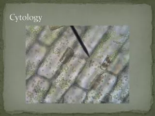
Cell membrane Trilamellar structure Dark (electron dense outer layer) Light (electron lucent middle layer)
Mitochondria rounded or oval, surrounded by two membranes; outer membrane is smooth and inner membrane forms shelves (cristae) Matrix shows electron dense granules: Ca+, RNA and DNA
Golgi apparatus Parallel flat membranous saccules transfer vesicles secretory vesicles
Lysosomes Primary: Homogenous rounded structure Secondary: 1- phagosome: rounded structure with heterogeneous matrix 2- multivesicular bodies: large vesicle with bibles in the matrix 3- autolysosome: old organelle inside (usually mitochondria)
Lysosomes Primary lysosome
Lysosomes Autolysosome