Download
1 / 34
510 likes | 1.51k Views
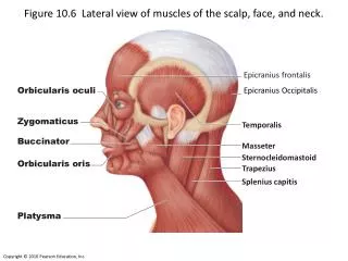
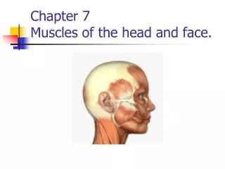
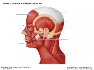
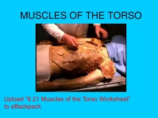
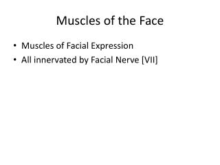
Muscles of the Face. Muscles of Facial Expression All innervated by Facial Nerve [VII]. Orbicularis Oculi. Functions to close the eyelids Palpebral part: closes eyelids gently Orbital part: closes eyelids forcefully. Levator Anguli Oris.

E N D
Muscles of the Face • Muscles of Facial Expression • All innervated by Facial Nerve [VII]
Orbicularis Oculi • Functions to close the eyelids • Palpebral part: closes eyelids gently • Orbital part: closes eyelids forcefully
LevatorAnguliOris • Raises corner of mouth; helps form nasolabial furrow
Depressor AnguliOris • Draws corner of mouth down and laterally
LevatorLabiiSuperioris • Raises upper lip; helps form nasolabial furrow
Depressor LabiiInferioris • Draws lower lip downward and laterally
Zygomaticus Major • Draws corner of mouth upward and laterally
Orbicularis Oris • Closes lips; protrudes lips
Occipitofrontalis • Frontal belly: wrinkles forehead; raises eyebrows • Occipital belly: Draws scalp backward
Buccinator • Presses the cheeks against teeth; compresses distended cheeks
Parotid Gland and Duct • Largest of 3 pairs of salivary glands in head • Facial nerve [VII], external carotid a., retromandibular v. pass through it. • Sensory innervation provided by auriculotemporal nerve (branch of mandibular nerve [V3]
Parotid gland Largest salivary gland Irregular shape Superficial & deep parts Tough fascial sheath Investing fascia Painful with “mumps” CV VII only passes through
Parotid duct Exits anterior surface of gland Accessory parotid along path of duct Crosses masseter Pierces buccinator Opens in oral vestibule opposite second upper molar
Parotid Bed (structures deep to parotid) 1. Mastoid process • Styloidprocess & three muscles 3. Stylomastoid foramen (exit of CN VII) 4. Posterior belly of digastric 5. Carotid sheath
Structures passing through parotid gland Facial nerve Retromandibular vein External carotid artery Superficial temporal and transverse facial Lymphatic vessels: superficial & deep parotid nodes (on sheath & within gland) Auriculotemporal nerve (carrying postganglionic parasympathetic fibers)
Innervation to Parotid Sensory: great auricular & auriculotemporal Vasomotor: sympathetic fibers via the external carotid plexus; nerve cell bodies are located in the superior cervical ganglion Secretion: parasympathetic fibers (GVEp) from CN IX
Innervation to Parotid CN IX Tympanic n Tympanic plexus Lesser petrosal Otic ganglion Auriculotemporal
Nerves of the Face • Supraorbital • Infraorbital • Mental • Auriculotemporal • Facial • Temporal • Zygomatic • Buccal • Mandibular • Cervical
Auriculotemporal Nerve • External acoustic meatus, surface of tympanic membrane, and large area of temple
Arteries and Veins of the Face Retromandibular Vein