Download
1 / 17
330 likes | 2.1k Views
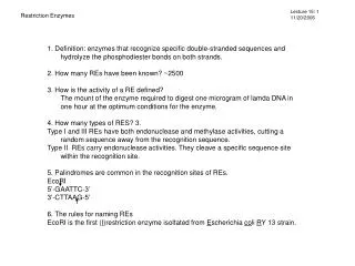
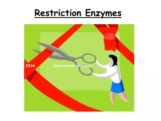
Restriction Endonucleases Or Restriction Enzymes. What Are Restriction Enzymes. They are enzymes that act as molecular scissors because they are able to cut DNA at specific base pairs. Each restriction enzyme recognizes a characteristic sequence of nucleotides, this is the recognition site.

E N D
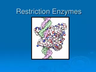
Restriction Endonucleases Or Restriction Enzymes
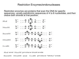
What Are Restriction Enzymes They are enzymes that act as molecular scissors because they are able to cut DNA at specific base pairs. Each restriction enzyme recognizes a characteristic sequence of nucleotides, this is the recognition site. The recognition site is 4-8 base pairs long and are usually characterized by a complementary palindromic sequence What makes a sequence palindromic is when both strands have the same base sequence when read in the 5’-3’ direction
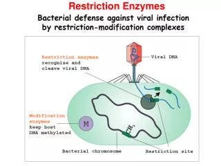
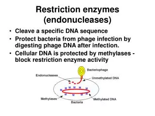
Where They Are Found Restriction enzymes are found in and extracted from single-celled bacterium. In the bacterium they serve as a protective mechanism against invading viruses. When a virus enters the bacterium, these enzymes are released and begin to cut the invading DNA into smaller harmless segments.
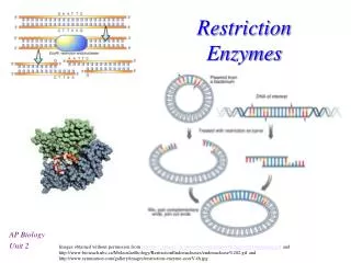
How It Works Restriction enzymes first expose the base sequence of a DNA fragment. They then scan for the recognition site to know where to cut the DNA strand. They then cut the phosphate backbones of the DNA molecules at the recognition site through a hydrolysis reaction. The hydrogen bonds of the complementary base pairs in between the cuts are disrupted.
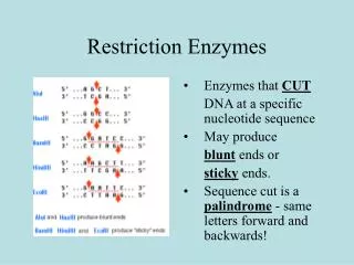
Making The Cut • The patterns of cuts varies with every restriction enzyme, however each enzyme will have its own manner of cutting, and will never change. • There are 3 different ways an enzyme can cut a sequence: • 5’ overhang: asymmetrical cutting, a short single-stranded piece of DNA overhangs from the 5’ end. BamHI is an enzyme that uses this way of cutting. • 3’ overhang: also asymmetrical cutting, a short single-stranded piece of DNA overhangs from the 3’ end. KpnI uses this way of cutting. • Blunts: this is when an enzyme cuts at precisely opposite sides of the 2 strands of DNA. It creates blunt ends without overhangs.
Sticky Ends Each single stranded fragment can pair with each other and join together When one strand of DNA extends beyond its complementary strand EcoRI results in sticky ends
Blunt Ends When the 2 complementary strands of DNA are of equal length Smal results in blunt ends
Methylases Restriction endonucleases have to be able to distinguish between foreign DNA, and that which is in their preexisting cells. If not the bacterium’s DNA could possibly be cleaved by the immune system. Methylases are specific enzymes found in both eukaryotes and prokaryotes. In prokaryotes they modify the recognition site of a respective restriction endonuclease by putting a methyl group on one of the bases, which prevents the resriction endonuclease from cutting the DNA into fragments. When foreign DNA is first brought into the bacterium it is not methylated, causing it to become defenceless against the bacterium’s restiction endonuclease. Methylases are important to molecular biologists when they work with prokaryotic organisms. They allow them to protect a gene fragment from being cleaved in an undesired location.
DNA Ligase If the genes are cut out, there has to be a way to put them back together. If 2 DNA fragments of nucleic acids have been generated using the same restriction enzyme, they are naturally attracted to each other at their complementary sticky ends. Hydrogen bonds form between the complementary base pairs, but this is not stable. The phosphodiester linkage between the backbones of the double strands has to be reformed. DNA ligase is the enzyme that is used for joining cut strands of DNA together. Using a condensation reaction, DNA ligase drives out a water molecule and helps reform the phosphodiester linkage. Joining strands with blunt ends using DNA ligase is inefficient. T4 DNA ligase is an enzyme that originated from the T4 bacteriophage, which joins blunt ends together.
Use In Biotechnology We use restriction enzymes in biotechnology to cut small strands of DNA to study the fragment length differences among individuals (Restriction Fragment Length Polymorphism) it’s also used in gene cloning. RFLP has been used to prove that each individual have distinctive differences in gene sequences and restriction cleavage patterns in certain areas of the genome. Using this knowledge we are able to use the method of DNA fingerprinting.
Types of Enzymes Type I: cuts DNA at random locations, can be as far as 1000 or more base-pairs from recognition site Type III: cuts at approximately 25 base pairs from the recognition site Type II: can generate 2 different types of cuts, depending on whether they cut both strands at the centre of the recognition sequence, or each strand closer to one end of the recognition sequence
Uses of Restriction Endonuclease • Helps provide a good immune system to fight off foreign DNA • Manipulation of E.coli bacteria to express recombinant human insulin for diabetes • It can also be used in criminal forensics. Forensic scientists use 13 regions of DNA to perform DNA fingerprinting. DNA is then isolated from the blood, saliva etc. The restriction enzymes are used to remove the 13 regions individually from the DNA to be fingerprinted, they are then isolated from the rest of the DNA. With the crime scene sample’s isolated DNA regions and the suspect DNA regions, the restriction enzymes are used again to chop DNA into shorter sections of varying lengths.
Restriction endonuclease animation http://highered.mcgraw-hill.com/olcweb/cgi/pluginpop.cgi?it=swf::535::535::/sites/dl/free/0072437316/120078/bio37.swf::Restriction%20Endonucleases http://www.phschool.com/science/biology_place/labbench/lab6/enzwork.html http://www.dnalc.org/resources/animations/restriction.html
Bibliography • Di Giuseppe, Maurice. Biology 12. Toronto. Thomson Nelson. 2003 • Phillips, Theresa. “Restriction Enzymes Explained”. http://biotech.about.com/od/proteinengineering/a/restrictenz.htm. About.com. 2010 • Roberts, Richard. “How restriction enzymes became the workhorses of molecular biology.” 2005. May 4 2010http://www.pnas.org/content/102/17/5905.ful • “Restriction Enzymes.” 23 December 2009. 5 May 2010. http://users.rcn.com/jkmball.ma.ultranet /BiologyPages/R/RestrictionEnzymes.htm • Tan, Alex. “Restriction Enzymes Used In Forensic Science”. http://www.ehow.com/facts_5724559_restriction-enzymes-used-forensic-science.html. eHow.1999-2010
QUIZ TIME!!! What type of restriction enzyme cuts at 25 base pairs from the recognition site? What is it called when one strand of DNA extends beyond its complementary strand? Name the 3 ways a restriction enzyme can cut a DNA sequence. What type of sequence has strands containing the same base sequence when read in the 5’-3’ direction?
QUIZ TIME!!! Where are restriction enzymes found? What enzyme helps rejoin cut DNA strands, that have produced sticky ends? What is the first step of how a restriction enzyme works?