Download
1 / 16
210 likes | 402 Views
Cerebrospinal fluid (CSF) analysis. Advanced laboratory techniques Third stage Lecture (6) ( Theortical ) Asisstant lecturer Rajaa Saihood Abbas. Cerebrospinal fluid (CSF) analysis.

E N D
Cerebrospinal fluid (CSF) analysis Advanced laboratory techniques Third stage Lecture (6) ( Theortical) Asisstant lecturer RajaaSaihood Abbas
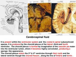
Cerebrospinal fluid (CSF) analysis Cerebrospinal fluid (CSF) analysis is a group of laboratory tests that measure chemicals in the fluid that surrounds and protects the brain and spinal cord. The tests may look for proteins, sugar (glucose), and other substances. CSF tests include the following : • CSF Physical Characteristics. • CSF Chemical Tests. • CSF Microscopic examination . • CSF infectious diseases tests .
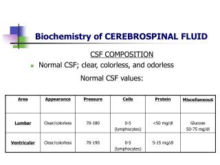
Physical Characteristics • The appearance of the sample of CSF is usually compared to a sample of water. • Pressure of the CSF can be measured when opening (starting) and finishing the collection. • Increased CSF pressure may be seen with a variety of conditions that increase pressure within the brain or skull and /or that obstruct the flow of CSF, such as tumors, infection, abnormal accumulation of CSF within the brain (hydrocephalus), or bleeding. • Decreased pressure may be due to dehydration, shock, or leakage of CSF through an opening (fracture).
Physical Characteristics • Colorof the fluid — normal is clear and colorless. Changes in the color of the CSF are not diagnostic but may point to additional substances in the fluid. Yellow, orange, or pink CSF may indicate the breakdown of blood cells due to bleeding into the CSF or the presence of bilirubin. Green CSF may also sometimes be seen with bilirubin or infection. • Turbidity — Cloudy or turbid CSF may indicate the presence of white or red blood cells, microorganisms, or an increase in protein levels. • Viscosity— Normal CSF will have the same consistency as water. CSF that is "thicker" may be seen in people with certain types of cancers or meningitis.
Chemical Tests • A few routine tests are usually performed on CSF samples. • CSF glucose – normal is about 2/3 the concentration of blood glucose. Glucose levels may decrease when cells that are not normally present use up (metabolize) the glucose. These may include bacteria or cells present due to inflammation (WBCs) or shed by tumors. •CSF protein – only a small amount is normally present in CSF because proteins are large molecules and do not cross the blood/brain barrier easily. Decreases in CSF protein are not generally considered significant. Increases in protein are most commonly seen with: • Meningitis and brain abscess. • Brain or spinal cord tumors. • Syphilis.
If any of the initial tests are abnormal or if the doctor has reason to suspect a specific condition, then additional testing may be ordered. This may include one or more of the following: 1- CSF protein electrophoresis — separates different types of protein. Oligoclonal bands may be seen with multiple sclerosis and Lyme disease. 2- CSF IgG (Immunoglobulin G) — increased in some conditions, such as multiple sclerosis, herpes encephalitis, connective tissue diseases. 3- Myelin basic protein — seen when the covering of nerves (myelin) breaks down, such as with multiple sclerosis. 4-CSF lactic acid — often used to distinguish between viral and bacterial meningitis. The level will usually be increased with bacterial and fungal meningitis while it will remain normal or only slightly elevated with viral meningitis.
5-CSF lactate dehydrogenase (LD) — used to differentiate between bacterial and viral meningitis; the level is usually increased with bacterial meningitis and not with viral meningitis; may also be elevated with leukemia or stroke. 6- CSF glutamine — may be increased with liver disease. 7- CSF C-reactive protein (CRP) is an acute phase reactant and is elevated with inflammation. It is markedly increased with bacterial meningitis. • Since it is very sensitive even with early bacterial meningitis, it is often used to distinguish between bacterial and viral meningitis. 8-Tumor markers — Carcinoembryonic antigen (CEA), alpha-fetoprotein (AFP), and HCG may be increased in cancers that have spread from other sites in the body (metastatic).
Microscopic Examination • Normal CSF has no or very few cells present and appears clear. If the CSF sample appears clear, a small drop of undiluted CSF is examined under a microscope, and cells are counted manually. If the number of cells present are very few (for example, 5 or less), the laboratory may or may not perform a cell differential . • If cells are numerous (such as greater than 5), a differential will most likely be done. To perform a differential, labs will often use a special centrifuge (cytocentrifuge) to concentrate the cells at the bottom of a test tube. A sample of the concentrated cells is placed on a slide, treated with special stain, and an evaluation of the different kinds of WBCs present is performed. • If cancer is suspected or has been previously diagnosed, the sample is usually cytocentrifuged regardless of the number of cells counted, and a differential is performed.
CSF total cell counts • Red blood cell (RBC) count. Normally no red blood cells are present in the CSF. The presence of red blood cells may indicate bleeding into the CSF or may indicate a "traumatic tap" - blood that leaked into the CSF sample during collection. • White blood cell (WBC) count. Normally less than 5 cells are present in the adult. A significant increase in white blood cells in the CSF is seen with infection or inflammation of the central nervous system.
CSF WBC differential test • Small numbers of lymphocytes, monocytes and, in neonates, neutrophil are normal in a sample of CSF. There may be: • An increase in neutrophils with a bacterial infection. • An increase in lymphocytes with a viral or fungal infection. • Sometimes an increase in eosinophils with a parasitic infection. • Abnormal and increased numbers of WBCs may be see with leukemia that is present in the central nervous system. • Abnormal cells may be present with cancerous tumors. • Immune disorders of the CSF, such as multiple sclerosis, may also cause a slight increase in lymphocytes.
CSF cytology • a cytocentrifuged sample is treated with a special stain and examined under a microscope for abnormal cells. This is often done when a central nervous system tumor or metastatic cancer is suspected. The presence of certain abnormal cells, such as tumor cells or immature blood cells, can indicate what type of cancer is involved.
Infectious Disease Tests • In addition to chemistry tests, such as protein and glucose, other routine tests may be performed to look for microorganisms if meningitis or encephalitis is suspected. • CSF gram stain for direct observation of microorganisms under a microscope. A sample of CSF is centrifuged and the concentrated portion is placed on a slide and treated with a special stain for examination under the microscope. There should be no microorganisms in CSF fluid. If bacteria or fungi are present on a CSF gram stain, then the patient has bacterial or fungal meningitis or encephalitis.
• CSF culture and sensitivity is used to detect any microorganisms, which will grow in the culture. If bacteria are present, they can be tested in the laboratory to predict the best choices for antimicrobial therapy for the affected person and prophylaxis (preventive treatment) of close contacts, if needed. If there are no microorganisms present, it does not rule out an infection; they may be present in small numbers or unable to grow in culture due to prior antibiotic therapy.
If any of the initial tests are abnormal or if the doctor strongly suspects a central nervous system infection, then additional testing may be ordered. This may include one or more of the following : • • Detection of viruses – detection of viral genetic material (DNA, RNA) by polymerase chain reaction (PCR) testing; for example, herpes virus and enteroviruses. Positive PCR tests for viral DNA or RNA, antigen tests, and growth on viral cultures indicate that a person has a viral infection and may have viral encephalitis or meningitis. • • CSF Cryptococcal antigen – to detect a fungal infection caused by the yeast Cryptococcus neoformans.
Specific CSF antibody tests – depending on which organism(s) are suspected. • Other CSF tests for infectious diseases that are less commonly ordered include: • CSF AFB smear and culture may be positive with tuberculosis and with other mycobacteria. • CSF molecular tests for Mycobacteria tuberculosis when tuberculosis is suspected. • CSF syphilis testing (VDRL) positive with neurosyphilis (involvement of the brain by syphilis) .


![CEREBRAL CIRCULATION AND CEREBROSPINAL FLUID [CSF]](https://cdn2.slideserve.com/4005143/slide1-dt.jpg)