Download
1 / 11
110 likes | 580 Views
Treatment of Microsporidial Keratitis with Hexamidine. Alex KH LAU 1 Colin SH TAN 1 Wee Jin HENG 1 1 Dept of Ophthalmology Tan Tock Seng Hospital, Singapore The authors have no financial interest in the subject matter of this e-poster. Introduction. Figure 1.

E N D
Treatment of Microsporidial Keratitis with Hexamidine Alex KH LAU1 Colin SH TAN1 Wee Jin HENG1 1Dept of Ophthalmology Tan Tock Seng Hospital, Singapore The authors have no financial interest in the subject matter of this e-poster.
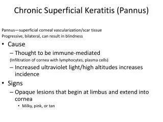
Introduction Figure 1 • Microsporidia are tiny, spore-forming, obligate intracellular eukaryotic protozoa (Figure 1). • In humans, microsporidia are opportunistic pathogens that usually cause diseases primarily in immunocompromised patients with Human Immunodeficiency Virus (HIV) infection. • 2 clinical entities of ocular microsporidiosis have been described1: • Corneal stromal keratitis in immunocompetent patients; caused by Nosema and Microsporidium and • Superficial punctate keratoconjunctivitis in immunocompromised individuals; mostly caused by Encephalitozoon.
Introduction • Treatment of ocular microsporidiosis is difficult and, to date, no definitive treatment exists. • Previously described treatment options include2-9: • Debridement, • Topical antibiotics (fluoroquinolones, propamidine isethionate, fumagillin), • Systemic anti-fungals & anti-helminths (itraconazole, albendazole) and • Topical steroids.
Objectives • We present a series of four cases of microsporidial keratoconjunctivitis in immunocompetent individuals who were treated successfully with topical hexamidine di-isethionate.
Case 1 • 48 year old Caucasian male • History of mud entered both eyes during rugby game in Cambodia. • Visual acuity was 6/12 in the right eye and intraocular pressure (IOP) was 34mmHg. Slit lamp examination revealed follicular conjunctivitis, multifocal subepithelial infiltrates, 2+ anterior chamber cells, as well as keratic precipitates on the endothelium (Figures 2 & 3). • Diagnosis of right microsporidial keratouveitis was confirmed with modified trichrome stain of corneal epithelial scrapings. • Patient was HIV negative. • He was treated with hexamidine di-isethionate, dexamethasone (preservative free), moxifloxacin and brimonidine. • Visual acuity recovered slowly to 6/6 over 3 months with minimal subepithelial scarring (Figure 4). Figure 2 Figure 3 Figure 4
Case 2 • 17 year old Chinese female • Developed redness and pain in the right eye 2 weeks after contact with mud. • Visual acuity was 6/6 bilaterally. There were multiple, coarse corneal epithelial & sub-epithelial infiltrates and follicular conjunctivitis (Figures 5 & 6). • Diagnosis of microsporidial keratitis was confirmed with modified trichrome stain. • She was treated with hexamidine di-isethionate, levofloxacin and oral albendazole. • The infection resolved over 3 weeks. Microsporidia keratitis subsequently developed in the left eye and was successfully treated with hexamidine di-isethionate. Figure 5 Figure 6
Case 3 • 23 year old Chinese male • Reported redness and pain in the left eye 4 days after contact with mud. • He was treated for viral conjunctivitis with topical tobramycin and dexamethasone. Five days later, he developed sub-epithelial infiltrates which worsened over the following week (Figures 7 & 8). • A clinical diagnosis of microsporidia keratitis was made and he was treated with topical hexamidine and levofloxacin. • Diagnosis was confirmed from corneal scrapings with modified trichrome stain. • The infection resolved over the next 2 weeks with no corneal scarring (Figure 9). • HIV status was negative. Figure 7 Figure 8 Figure 9
Case 4 • 63 year old Chinese male • Presented with redness, blurring of vision, pain, photophobia and discharge in the right eye. • VA was 6/7.5 and diffuse subepithelial infiltrates were seen with few keratic precipitates. • Clinical diagnosis of microsporidial keratitis was made • He was started on hexamidine di-isethionate and the keratitis resolved completely after 6 weeks of treatment.
Discussion • Our case series reports the successful treatment of ocular microsporidiosis with hexamidine and its manifestations in healthy, immunocompetent individuals. • There are increasing reports describing ocular microsporidiosis in healthy individuals. This could be explained by the increased awareness of this rare infection and improvement in the diagnostic techniques10, 11. • Our first case presented with a moderately severe uveitic response in the anterior chamber, which has not previously been reported. This could be due to sterile inflammatory reaction, or may represent a new clinical manifestation of ocular microsporidiosis. • Three of the 4 cases in our series developed microsporidial keratitis after exposure to mud, which is consistent with previous reports of trauma as one of the predisposing factors for ocular microsporidiosis in healthy individuals. The others include topical steroid therapy (Case 3) and contact lens wear.
Conclusion • Microsporidia are increasingly become recognised as pathogens in healthy individuals. • High index of suspicion is required to make the correct diagnosis, especially in cases presented with atypical multifocal diffuse epithelial keratitis. • History of ocular trauma, contact lens wear or usage of topical steroid therapy are predisposing factors which should raise the index of suspicion. • Topical hexamidine di-isethionate is an effective alternative therapy to microsporidial keratoconjunctivitis.
References 1 Weber R, Bryan RT, Schwartz DA, Owen RL. Human microsporidial infections. Clin Microbiol Rev 1994;7:426–61. 2 Metcalfe TW, Doran RM, Rowlands PL, et al. Microsporidial keratoconjunctivitis in a patient with AIDS. Br J Ophthalmol 1992;76:177– 8. 3 Friedberg DN, Stenson SM, Orenstein JM, et al. Microsporidial keratoconjunctivitis in acquired immunodeficiency syndrome. Arch Ophthalmol 1990;108:504–8. 4 Yee RW, Tio FO, Martinez JA, et al. Resolution of microsporidial epithelial keratopathy in a patient with AIDS. Ophthalmology 1991;98:196 –201. 5 Diesenhouse MC, Wilson LA, Corrent GF, et al. Treatment of microsporidial keratoconjunctivitis with topical fumagillin. Am J Ophthalmol 1993;115:293– 8. 6 Rosberger DF, Serdarevic ON, Erlandson RA, et al. Successful treatment of microsporidial keratoconjunctivitis with topical fumagillin in a patient with AIDS. Cornea 1993;12:261–5. 7 Gritz DC, Holsclaw DS, Neger RE, et al. Ocular and sinus microsporidial infection cured with systemic albendazole. Am J Ophthalmol 1997;124:241–3. 8 Theng J, Chan C, Ling ML, et al. Microsporidial keratoconjunctivitis in a healthy contact lens wearer without human immunodeficiency virus infection. Ophthalmology 2001;108:976–8. 9 Rossi P, Urbani C, Donelli G, Pozio E. Resolution of microsporidial sinusitis and keratoconjunctivitis by itraconazole treatment. Am J Ophthalmol 1999;127:210-2. 10 Davis RM, Font RL, Keisler MS, Shadduck JA. Corneal microsporidiosis. A case report including ultrastructural observations. Ophthalmology 1990;97:953–7. 11 Silverstein BE, Cunningham ET Jr, Margolis TP, et al. Microsporidial keratoconjunctivitis in a patient without human immunodeficiency virus infection. Am J Ophthalmol 1997;124:395– 6.