Download
1 / 18
180 likes | 245 Views
Microbial keratitis diagnosis and management, including initial investigation, treatment phases, antibiotic therapy, corneal culture, and sterilization approaches. Learn about the importance of antibiotics, culture materials, and treatment strategies for this eye condition.

E N D
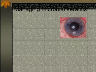
Diagnosis Microbial keratitis is rare in the absence of predisposing factors which include (1) contact lens wear, (2) ocular surface disease, eg (a) herpetic keratitis, (b) corneal-anaesthesia, (c) exposure and (d) bullous keratopathy and (3) trauma. Infection can present as an epithelial defect or corneal melt, without inflammation, in the immunocompromised or in those using topical steroids in whom corneal infiltration by leucocytes, the hallmark of microbial keratitis, may be absent.
Initial Investigation • Corneal scraping is indicated whenever microbial keratitis is suspected. It provides material for a microbiological diagnosis, debrides necrotic tissue and enhances antibiotic penetration. Endophthalmitis does not follow bacterial keratitis without corneal perforation (unlike fungal keratitis) so that anterior chamber and vitreous taps are not indicated when perforation is absent
Corneal culture materials • Corneal culture materials should be available in the emergency area and include, as a minimum, a slide for Gram staining and a blood agar plate for aerobic incubation. Most corneal isolates in temperate areas, including fungi, will grow on these media. Ocular specimens should be inoculated directly onto the media avoiding the use of transport or storage media. Some of the pathogens isolated from ocular infections may be considered to be normal flora by non-ocular microbiologists, so it is important that ophthalmologists liaise closely with the laboratory. A 21 gauge needle may be used to take the specimens.
Treatment • Therapy can be divided into a sterilisation phase and a healing phase with clearly defined endpoints for clinical review and decision making. Clinical signs may not indicate when corneal sterilisation has occurred, after starting intensive therapy, because sterilisation often precedes both epithelial healing and the resolution of inflammatory signs.
Treatment • These may also be delayed by preservative or agent related toxicity where intensive topical treatment is prolonged. Intensive antibiotic therapy is given for a limited period in the stetilisation phase and is followed by the healing phase, in which reduced therapy is aimed at (1) limiting further inflammatory damage, (2) preventing superinfection, and (3) promoting epithelial healing.
Treatment • Antibiotics should consist of broad spectrum topical antibacterial treatment because (1) a negative Gram stain does not exclude keratitis, (2) bacterial isolates are far more common than fungi or amoebae and (3) polymicrobial infections are common. This approach is continued unless the infecting agents are identified, with their antimicrobial sensitivities, enabling specific therapy to begin.
Antibiotics • The choice of antibiotics now hes between the standard regimen of topical, commercially unavailable, fortified aminoglycoside and fortified cephalosporin drops (ie gentamicin 1.5% and cefuroxime 5%) or the new regime of fluoroquinolone monotherapy with commercially available ciprofloxacin or ofloxacin 0.3%.
Broad spectrum cover against the majority of bacterial pathogens • Both the standard and new regimens offer broad spectrum cover against the majority of bacterial pathogens but fluoroquinolone monotherapy has advantages and has been shown to have equal efficacy in recent clinical trials. • However, fluoroquinolones may not adequately treat streptococcal keratitis and comination therapy of a quinolone with a fortified cephalosporin may be advisable in patients with ocular surface disease or in children in whom streptococcal infection is more common. • Currently both the standard and fluoroquinolone regimen encounter bacteriola resistance in about 5% of cases. • Neither regimen treats fungal or acanthamoeba infection.
Sterilisation Phase • Hourly administration of topical antibiotic therapy for five days leaves a wide rnargin of safety for most bacterial infections and compares well with a gradual reduction of high dose antibiotic treatment. Most cases can be managed successfully as outpatients with initial review after forty eight hours. Admission may be necessary where good compliance is unlikely or for overnight treatment in severe infections (axial lesions, lesions 6mm or more in diameter, or with 50%, or more stromal thinning). Systemic antibiotics (ie ciprofloxacin 750mg bd) ire indicated where the ulcer is close to the limbus. This may help protect from contiguous spread of infection to the sclera and enhance antibiotic delivery to peripheral lesions. Adjunctive treatment at this stige may include (1) dilating drops, (2) analgesic medication or (3) hypotensive agents for secondary glaucoma. Subconjunctival injections should be avoided.
Sterilisation Phase • Amoebic and fungal kerititis are rarely rapidly progressive and may be exacerbated by bacterial superinfection. Specific investigations and treatment regimens, involving a much more prolonged sterilisation phase, are required for both and lie outside the scope of this review. However, unless there is clear clinical evidence (or a Gram stain) suggesting non-bacteriid infection, it is appropriate to commence treatment with intensive broad spectrum antiibiotics for a defined initial period.
Interpretation of Culture Whilst cultures should be incubated for a minimum of 14 days before being reported as culture negative, growth of most pathogens can be expected ifter 48 hours. • Sensitivity Testing • If clinical progress is satisfactory, there is no indication for altering antibiotic therapy. But sensitivity testing can be invaluable in guiding the choice of a more appropriate antibiotic where bacterial keratitis is progressive. • Review at one week is necessary to determine whether the disease is progressive or resolving. Clear evidence of poor compliance or, in culture positive cases, resistance to the initial antibiotic choice are indications for re-entering the sterilization phase using appropriate specific therapy. Deteriorating or static cases should be referred, whereas cases in which resolution is partial but incomplete may safely enter a second phase of treatment directed at encouraging healing.
Initial Review • Early review at two days is to detect rapidly progressive cases and assess the culture results. Daily review can be confusing as the inflammatory reaction may be enhanced by endotoxin release within the first 48 hours. • However definite progression at this stage (increased stromal thinning or a clear expansion of the ulcer) is unusual, and implies that patients are either insensitive to, or not complying with, antimicrobial therapy. • This rapid early progression can be treated by admitting patients to ensure compliance and reviewing the microbiology results. Unless these indicate resistance to the primary therapy, with a change to a more appropriate antibiotic, then continue initial broad spectrum antibiotic therapy until two days of hourly treatment day and night have been followed by a further three days of hourly treatment during the day. Further progression after this point is then an indication for specialist referral. • Threatened or actual perforation indicate urgent referral as emergency penetrating keritoplasties in these circumstances carry a poor prognosis for vision, are difficult to perform well, and can often be avoided even after a perforation.
Healing Phase Healing is commonly retarded by persisting inflammation, treatment toxicity or untreated underlying ocular surface disease. Antibiotic treatment should be reduced to prophylactic levels at this stage to avoid toxicity and unpreserved medication used where possible. • Ocular surface disorders (ie dry eyes, exposure, entropion and blepharitis) must be treated. Complete resolution of anterior chamber and comeal inflammatory signs is normal in microbial keratitis without steroid treatment. However, topical steroids may speed re-epithelisation and resolution of the inflammatory response although their use will enhance fungal or herpetic infection and may increase the risk of perforation by inhibiting wound healing. In corneal graft recipients, without evidence of fungal infection, steroid therapy should be introduced at the outset to protect against a rejection episode. • At review after one week of the healing phase (ie week 3 after presentation), referral may be indicated as indolent ulceration may be due to unusual organisms, usually of low virulence, with continued disease progression