Diffuse Lamellar Keratitis
Diffuse Lamellar Keratitis. Eric E. Polk, OD, FAAO TLC Laser Eye Center St. Louis, MO. Diffuse Lamellar Keratitis (DLK) Sands of Sahara Syndrome.

Diffuse Lamellar Keratitis
E N D
Presentation Transcript
Diffuse Lamellar Keratitis Eric E. Polk, OD, FAAO TLC Laser Eye Center St. Louis, MO
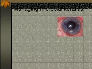
Diffuse Lamellar Keratitis (DLK) Sands of Sahara Syndrome • DLK is a non-infectious, inflammatory condition associated with LASIK. It is characterized by a sterile infiltration of inflammatory cells along the flap-stroma interface. It can vary from a mild self-limiting condition to a severe condition associated with stromal melting and decreased best corrected vision.
Etiology of DLKDLK was first described in the literature in 1998. • Interface Debris • Fibers • Red blood cells • Meibomian cells • Glove talc • Microkeratome oil
Risk factors for Developing DLK • Microkeratome induced epithelial defects • Corneal abrasions • Recurrent erosions • Iritis • Viral keratoconjunctivitis
Sporadic DLK • DLK developing in 1 or 2 patients a day. • Typically less severe. • Sporadic DLK after LASIK. Steve Wilson, MD. Cornea, 2001:21:560-3 • Exaggerated cellular response. • Dr. Wilson theorizes that sporadic DLK has a different etiology then epidemic DLK
Epidemic DLK • Occurs in 3 or more patients in one surgery day. • Epidemic DLK (cluster) is a more severe form of DLK. • Likely initiated by a causative agent. • Surgical gloves: silicone oil • Microkeratome blades • Contaminated sponges • Bacterial biofilm releasing endotoxins
DLK Etiology- Allergies • Boorstein. Atopy: A patient-specific risk factor for DLK, Ophthalmology 2003. • Found that DLK is more prominent in individuals who have untreated atopy. • Patients who took a histamine receptor 1 antagonist for allergies did not have an increased risk • We recommend keeping patients on their allergy medications before surgery.
Identification of Stage 1 DLK • It is imperative to identify DLK in its beginning stages. • Subtle appearance in early stages • Typically presents the first day after surgery.
Identification of Stage 1 DLK • Cells present at peripheral flap margin • Originate from limbal blood vessels • 120 micrometers below anterior surface • White blood cells • Diffuse scattering of cells • Cells are uniform in size
Differential Diagnosis • Surgical Debris • Meibomian Cells • Interface Haze • Infectious Infiltrates • Difference • Debris remains stationary • DLK moves centrally
Stages of DLK • Stage 1 • Stage 2 • Stage 3 • Stage 4 • Linebarger, Hardten, Lindstrom. J Cataract Refract Surg. 2000 Jul;26(7):1072-7.
Stage 1 • 1 in 50 cases • Cells present in the peripheral flap • Begins 1st day after surgery or injury • Patients are asymptomatic • Stage 1 DLK Categorization System • Area of interface effected • Mild Density • Moderate Density • Severe Density
Stage 2 DLK • 1 in 200 Cases. • Cells move to center of axis. • Mild, moderate, severe density • 2-4 Days after surgery. • May begin to have vision distortion.
Stage 3 DLK • 1 in 500 Cases • Cells clump and aggregate in central cornea • More likely to develop visual symptoms • 3-5 days after surgery • Collagenase enzymatic reaction starts • Requires immediate wash out of interface
Stage 3 Stage 4
Stage 4 DLK • 1 in 5000 Cases • 4-9 Days Post-Op • Stromal degradation and flap melt • Irregular astigmatism • Loss of BCVA
Time Table For DLK • Stage 1: Day 1-2 • Stage 2: Day 2-4 • Stage 3: Day 3-5 • Stage 4: Day 4-9 • Routine check 2-4 days • DLK patients 1-2 days
Treatment of Stage 1 DLK • Topical prednisilone q 1-2 Hrs • Oral Prednisone 60-80 mg • For moderate to severe cases • Keep antibiotic the same • Follow Up 1-2 Days
Oral Prednisone • Write RX in 10 mg tablets • 20 mg BID for smaller/ normal size individuals • 30 mg BID for larger persons • May cause insomnia or nightmares
Treatment of Stage 2 DLK • Requires topical prednisilone every hour. • Start oral steroids if not already started. • See patient next day. • Include surgeon in decision making process.
Treatment of Stage 3 DLK • Lift flap and irrigate interface • Wash-out must be done ASAP • Almost always very effective.
DLK secondary to trauma • Harrison. DLK associated with recurrent corneal erosions after LASIK, J Refract Surg 2001;17:463-65 • Haw. DLK associated with an epithelial defect in six eyes, J Refract Surg 2000; 16: 744-48. • Keszei. DLK associated with iritis 10 months after LASIK, J Cataract Refract Surg 2001; 27: 1126-27.
Secondary DLK • Consider treating corneal abrasions with prophylactic topical steroid.
Secondary DLK • DLK may occur in patients months to years after LASIK surgery • Report of DLK up to 3 years after LASIK surgery • Jin GJ, Lyle WA, Merkley KH. Late-onset idiopathic diffuse lamellar keratitis after laser in situ keratomileusis. J Cataract Refract Surg. 2005 Feb;31(2):435-7.
IOP Induced Stromal Keratitis(PISK) • Fluid accumulation in the interface secondary to corticosteroid induced elevated intraocular pressures. • Very unusual • Occurs after prolonged, or intensive use of corticosteroids. • 5% of population are steroid responders. • Appears clinically identical to severe DLK.
3 Day LASIK Post-Op • 20/20 BCVA • Eye is white and quiet • Diagnosis and Treatment
3 Day LASIK Post-Op • 20/20- BCVA • Eye is white and quiet • Diagnosis and Treatment
1 Month PRK Post-OP • Meds: FML QID x 1 Month • BCVA: 20/25 • Conj: White • A/C Clear • Diagnosis and Treatment
Interface • Definition- The interface is an area that is created by a microkeratome during LASIK surgery. It is the area between the flap and stromal tissue. It is unique to LASIK patients.
3 Day Post-op LASIK • Hyperemic Conj • +1 A/C Reaction • Symptomatic • Pain • Photophobia
1 Day Post-Op LASIK • 20/20 BCVA • Conj is white • Mild discomfort • Diagnosis and Treatment
5 Day LASIK Post-Op • BCVA: 20/50 • Conj: White • A/C: Clear • Diagnosis and Treatment?