
Functional Genomics Clearinghouse from Hiram College
Discover how undergraduate and high school students explore functional genomics through genetics and biology courses at Hiram College. Learn about their efforts in genome sequencing, mutant analysis, and more.

Functional Genomics Clearinghouse from Hiram College
E N D
Presentation Transcript
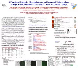
Hiram genomics academy A Functional Genomics Clearinghouse as an Outcome of Undergraduate& High School Education – An Update of Efforts at Hiram CollegeBrad Goodner1, Cathy Wheeler1, Prudy Hall1, Stuart Gordon1, Kathryn Reynolds1, Stephanie Lammlein2, Lindsey Wilson1, Garrett Temkiewicz1, 2002-2007 Molecular & Cellular Biology courses1, 2002-2007 Genetics courses1, 2006-2007 Introductory Biology courses2, & 2006-2007 Hiram Genomics Academy sessions31 Hiram College, Hiram, OH (goodner@hiram.edu, 330-569-5260), 2 Rootstown High School, Rootstown, OH,& 3 81 high school students from all over Ohio, Michigan, Pennsylvania, & Indiana Abstract Obtaining the complete genome sequence of any organism is really just a new beginning – you have a lot of tools now available but are not yet sure how best to use them. Functional genomics is really just an extension of how molecular biology and microbiology have made progress over the past 25 years or more through characterization of mutants through forward or reverse genetics. The extension is one of breadth to include as many genes as possible. Over the past 6 years, Hiram College faculty and students have accumulated a large set of mutations in the A. tumefaciens C58 genome (and to a lesser extent in the A. rhizogenes A4 genome). Students in the Molecular & Cellular Biology course use reverse genetics to test functional predictions based on bioinformatics analyses of gene function. We will highlight the work done by the 2006 and 2007 iterations of the course. Students in the Genetics course, along with high school students during the past two years, have used forward genetics to link genes to functions through a large scale mutant hunt. We will highlight some of the more interesting findings in several functional categories. Finally, we will look forward to future efforts and we are certainly open to new suggestions from others. 2007 Molecular & Cellular Biology Course – Penicillin Binding Proteins The bacterial cell wall is crucial for withstanding turgor pressure and also as a cell shape determinant. The wall is composed of a heteropolymer called peptidoglycan that has both polysaccharide and peptide characteristics. The major enzymes involved in wall synthesis, maintenance, and turnover are nicknamed Penicillin-Binding Proteins (PBP’s) because the antibiotic penicillin and its derivatives act as irreversible enzyme poisons on these proteins. Most rod-shaped bacteria have 6 or more PBPs that fall into 4 categories. Class A PBP’s construct new peptidoglycan chains and crosslink them. Class B PBP’s are only involved in cros-linking peptidoglycan chains, but do so in either a elongation-growth or septation-growth mode depending on the enzyme. The low molecular weight PBP’s modify peptidoglycan chains before or after crosslinking. Finally, cell wall-localized b-lactamases, besides their impact on antibiotic resistance, can be involved in peptidoglycan turnover. E. coli and Bacillus subtilis have been the models for studying PBP activities. Data from these two organisms have shown that the Class B PBP’s influence cell shape in terms of the lengths of rods or rod-to-coccus transitions because of their differential activities during the cell cycle, and the low MW PBP’s influence the uniformity of cell shape by altering the cross-linking level and substrate availability. The Fall 2007 Molecular & Cellular Biology course at Hiram College (50 students) set out to look at the function of the 11 PBP’s encoded by the A. tumefaciens C58 genome. During the course of a 12-week semester, the students were able to make mutations in 9 of the 11 genes and to obtain phenotypic data on 8 of the mutants. Here are their initial observations (also see Table 1): * While some of the mutants might have a slight change in cell shape, the Atu1341- mutant had a dramatic cell shape change and showed a large decrease in growth rate. The Atu1341- cells had a variety of amorphous shapes with few regular rods. * Most of the mutants had a definite impact on cell motility. The Atu1499- mutant is very hypermotile, as compared to wildtype, on both 0.3% and 0.9% agar plates. The Atu1341- mutant is hypermotile only on 0.3% agar. The Atu1067-, Atu1505-, Atu2513-, and Atu3694- mutants were hypomotile on all agar concentrations tested. Summary of Accomplishments Up to Date Reverse Genetics 75 specific genes disrupted so far & mutant phenotypes characterized Forward Genetics ~10,000 Agrobacterium transposon insertion mutants generated ~10,000 mutants screened for 10 different phenotypes 160 mutants with interesting phenotypes saved for further analysis genomic DNA isolated from 119 of these mutants the site of transposon insertion cloned out & sequenced Interesting Stories from Forward Genetics Screen for Biofilm Defects (change in colony morphology on LB agar minus NaCl plus Coomassie Blue and Congo Red; Figure 2): Atu2121 on ChrI encodes Lyc glycosyl hydrolase may be involved in exopolysaccharide synthesis or maintenance Atu2660 on ChrI (HCA61) encodes a conserved hypothetical protein Atu3327 on ChrII (HCA41) encodes ExoY succinoglycan exopolysaccharide synthesis protein Atu3437 on ChrII encodes a ABC transporter ATP-binding protein Atu3740 on ChrII (HCA82) encodes fructose bisphosphate aldolase may influence substrate availability for making exopolysaccharide Atu4000 on ChrII (HCA40) encodes BioA adenosylmethionine-8-amino-7-oxononanoate aminotransferase role of biotin in biofilm? – in E. coli, bioF- mutant has altered biofilm Atu4053 on ChrII encodes ExoA succinoglycan biosynthesis protein Atu4668 on ChrII encodes a ABC transporter permease Screen for pH Tolerance (lack of growth on LB agar pH 5.5 or LB 10): 15 acid sensitive mutants all of them only show defect on solid medium! Why? 4 base sensitive mutants (Figure 3) most interesting is Atu0512 encoding PhaA pH adaptation K+ efflux system component mutation in Atu0512 homolog of Sinorhizobium is base-sensitive & is nodulation-negative experiments underway to see if Atu0152- mutant is virulent Screen for Auxotrophy: identified steps in pathways for aspartate, branched chain amino acid, histidine, methioinine, purine, pyrimidine, serine & glycine, & tryptophan synthesis mutations in glucose dehydrogenase & transaldolase point out crucial role for Entner- Doudoroff & pentose phosphate pathways mutation in glycogen synthase impacts use of glucose but not of glycerol mutation in Atu3885 encodes a inositol monophosphatase family member that is needed for growth on both glucose and glycerol; role? mutation in Atu1457 leads to auxotrophy for sulfur; gene encodes a conserved hypothetical protein ; in many organisms, this gene is right next to Atu1456 encoding CysI sulfite reductase hemoprotein beta subunit (Figure 4); our working hypothesis is that Atu1457 encodes a previously unknown protein of the cysteine synthesis pathway; students at Rootstown High School are currently working to see if the auxotrophic phenotype in Atu1457- mutant is due to that mutation or just a polar effect within the operon A B 2006 Molecular & Cellular Biology Course – 24 Two-Component System Mutants, 12 Environmental Variables, 54 Treatments Two-component systems are a common regulatory mechanism in bacteria and the A. tumefaciens genome is loaded with 34 paired teams, 5 hybrid proteins, 17 orphan histidine sensor kinases, and 16 orphan response regulators. Previous work by others had characterized 7 proteins that fall into this family. The 2005 and 2006 iterations of the MolCell course knocked out 11 and 16, respectively, of the response regulator genes. The 2006 course used a new microtiter plate reader to study the phenotype of 20 of the response regulator mutants. A summary of their findings is given in Table 2. C Figure 2. Biofilm defective mutants. A: Colony morphology of selected mutants on LB-NaCl+dyes. Wildtype is shown at the top. B: Pellicle morphology of selected mutants. Wildtype is shown at the top. C: Assay for binding to polystyrene microtiter wells under different [Pi]. Table 2. Summary results for a phenotypic microarray of 20 response regulator mutants. Mutant StrainCondition* Atu0050- (actR)glucose-low,glucose, sucrose, xylose, succinate,ammonium-low, nitrate, glutamine, arginine, 1%NaCl, 2%NaCl, 1.5%KCl, 2%NaCl, 3%KCl,Mg-both5.5, Mg-low7, temp-35C, pH10, Ca-all Atu0114- (dctD)succinate, 2%NaCl, Mg-both5.5, pH5.5, Ca-0.1mM, Ca-none,0.1uM,10mM Atu0343- (barA)succinate, Atu0527- glucose-low,succinate, 2%lactate Atu0629-pH5.5 Atu0970- (phoP)succinate, 3%KCl, Mg-both5.5, pH5.5, temp-15C Atu0978- (ragA)glucose-low,Mg-both5.5, pH5.5,Fe-DIP Atu1116- (nwsB)glucose-low, succinate,Mg-high5.5, pH5.5 Atu1297- (pleD)proline,2%NaCl Atu1446- (ntrC)glucose-low, succinate, serine(C), 2%NaCl, 3%KCl,Fe-M9 Atu1448- (ntrX)succinate, 2%NaCl, pH5.5 Atu1900- (agp1)glucose, glucose-low, sucrose, xylose, glycerol, succinate, histidine(C), serine(C), ammonium-low, nitrate, proline, 1%NaCl, 1.5%KCl, 2%NaCl, 3%KCl,Mg-both7, 1%lactate, 2%lactate, 1%urea, 2%urea, temp-all, pH5.5, phosphate-all, Ca-all, Fe-all, H2O2, Cu Atu2165- (agp2)glucose, glucose-low, sucrose, xylose,glycerol, succinate, histidine(C), serine(C), ammonium-low, nitrate, serine, proline, 1%NaCl, 1.5%KCl, 2%NaCl, 3%KCl, Mg-both7, 1%lactate, 2%lactate, 1%urea, 2%urea, temp-all, pHall, pH10, phosphate-all, Fe-all, H2O2, Cu, Oxyrase, anaerobic Atu2206- (petR)1.5%KCl, 3%KCl Atu2332- succinate Atu2434- (ctrA)succinate, 2%NaCl, Mg-high5.5 Atu3907- (nasT)2%NaCl, temp-15C Atu4047- (cvgS)succinate,proline,Mg-both5.5, pH5.5 Atu4300- (nwsB)glucose, sucrose, xylose, histidine(C), serine(C), nitrate, 1%NaCl, 2%NaCl, 1.5%KCl, 2%NaCl, 3%KCl,Mg-both5.5, Mg-low7, 1%lactate, 2%lactate, 1%urea, 2%urea, pH10 Atu4805-xylose, 2%lactate, 2%urea, Ca-10mM * Interpretation of impact results: blue means the mutant showed growth >50% greater than that seen for wt red means the mutant showed >50% less growth than that seen for wt bold means in a ratio to wt under that condition (e.g., mutant at pH5.5/wt at pH5.5) underlined means as a ratio to itself at baseline condition (mutant pH5.5/mutant pH7 compared to wt 5.5/wt 7) Figure 3. Atu0512- mutant is base-sensitive. Growth curve of Atu0512- mutant in LB broth at different pH levels. Table 1. Data on PBP- mutants obtained by 2007 Molecular & Cellular Biology course. Gene Current Best Hit Domains identified Possibly Growth Impact on NumberAnnotationto E. coliby InterProScan*EssentialDefectMotility Class A PBP’s (have both glycosyltransferase & transpeptidase activities): Atu0103 PBP1A PBP1B 1,2,3,4,14 N N Hypomotile Atu0931 PBP PBP1B 1,2,3,4 N N N Atu1341 PBP1A PBP1A 1,2,3,4 N Very slow Hypermotile Atu3694 PBP PBP1C 1,2,4,12,13 N N Hypomotile Class B PBP’s (have only transpeptidase activity): Atu1067 PBP PBP2/3 2,8,9 N N Hypomotile Atu2100 PBP2 PBP3 2,4,8,9 N no data no data Low MW PBP’s (have either DD-carboxypeptidase or endopeptidase activity): Atu1499 Dac PBP5/6 4,5,6,7 N Slow Very hypermotile Atu1505 Dac PBP5/6 2,6,10 N N Hypomotile Atu2321 Dac PBP5/6 2,6,10 Y? Atu3634 DacF PBP5/6 2,6 Y? b-Lactamases: Atu2513 PBP b-lactamase 11 N N N * InterProScan Domains: 1 = Family 51 glycosyl transferase domain 2 = PBP transpeptidase domain 3 = PBP1a domain 4 = Beta-lactamase transpeptidase domain 5 = Peptidase S13 D-Ala-D-Ala carboxypeptidase C domains 6 = Peptidase S11 D-Ala-D-Ala carboxypeptidase A domains 7 = PBP-associated domain 8 = Class A Beta-lactamase 9 = PBP dimerization domain 10 = Cell division protein domain 11 = Beta-lactamase family 12 = PBP1c domain 13 = PBP C-terminal domain 14 = WW/Rs5/WWP domain Atu1456Atu1457 Atu1456Atu1457 A B C Figure 1. Cell shape of the Atu 1341- mutant is highly altered. A, B: Atu1341- mutant. C, D: Atu1499- mutant. E, F: Atu2513- mutant. A, C, E: Light microscopy of crystal violet-stained cells. B, D, F: Fluorescent confocal microscopy of Bocillin FL-labeled cells (fluorescent penicillin derivative). Figure 4. Atu1457 orthologs are in a consistent genomic context. A: Position of the Atu1457 ortholog (pale green arrow) just behind Atu1456 CysI ortholog (bright red arrow) in a wide variety of genera within the alpha-Proteobacteria. B, C: Similar alignment for members of the gamma-Proteobacteria and beta-Proteobacteria, respectively. Atu1456 CysI ortholog shown in pale green while Atu1457 ortholog shown in bright red. This work was funded by Hiram College and by A B C D E F


