Download
1 / 52
560 likes | 990 Views
The Neuromuscular Junction And Muscle Stimulation. Skeletal Muscle Fibers. A muscle fiber is a multinucleated muscle cell that attaches to connective tissue. Sarcolemma is the muscle cell membrane. Sarcoplasm is the cytoplasm containing nuclei, mitochondria, and myofibrils.

E N D
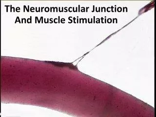
The Neuromuscular Junction And Muscle Stimulation
Skeletal Muscle Fibers • A muscle fiber is a multinucleated muscle cell that attaches to connective tissue. • Sarcolemma is the muscle cell membrane. • Sarcoplasm is the cytoplasm containing nuclei, mitochondria, and myofibrils. • Myofibrils are composed of protein filaments, predominantly myosin and actin.
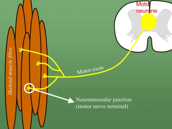
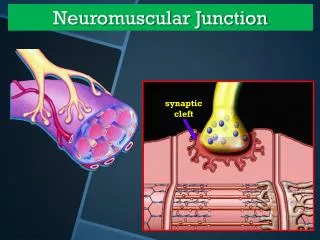
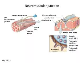
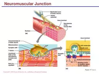
Motor neuron axons join the skeletal muscle at the neuromuscular junction. • Motor end plate is a specialized region of the sarcolemma at the neuromuscular junction.
A motor unit consists of the motor neuron and the muscle fibers it controls.
Synaptic cleft is a space between the neuron and the motor end plate.
Neurotransmitters are chemicals stored in vesicles of the motor neuron axon. Acetylcholine (ACh)controls skeletal muscle contraction.
Intracellular Structure of Muscle • Sarcoplasmic reticulum: network of membranous sacs surrounding myofibrils. (Contains Calcium) • Transverse tubules (T-tubules) extend deep into the sarcoplasm and contain extracellular fluid. These transverse tubules allow a multinucleated muscle fiber to be stimulated simultaneously. • Cisternae: enlarged portions of the sarcoplasmic reticulum. (Contains Calcium)
Sarcoplasmic Reticulum (SR) • SR is an elaborate, smooth endoplasmic reticulum • runs longitudinally and surrounds each myofibril • Form chambers called terminal cisternae on either side of the T-tubules • A single T-tubule and the 2 terminal cisternae form a triad • SR stores Ca++ when muscle not contracting • When stimulated, calcium released into sarcoplasm • SR membrane has Ca++ pumps that function to pump Ca++ out of the sarcoplasm back into the SR after contraction
Muscle Contraction Summary • Nerve impulse reaches neuromuscular junction • Acetylcholine is released from motor neuron • Ach binds with receptors in the muscle membrane to generate signal in the sarcolemma
Muscle Contraction (Cont’d) • Activation signal travels down T tubule • Sarcoplamic reticulum releases calcium • Calcium binds with troponin to move the troponin, tropomyosin complex • Binding sites in the actin filament are exposed
Muscle Contraction (cont’d) • Myosin head attach to binding sites and create a power stroke • ATP detaches myosin heads and energizes them for another contraction • When activation signals cease the muscle stops contracting
Events of Muscle Relaxation • Acetylcholine is degraded by the enzyme acetylcholine-esterase (AChE)and the muscle is no longer stimulated. • Calcium ions are actively transported back into the SR. • Actin-myosin linkages break. • Troponin and tropomyosin cross-bridges reform. • Troponin and tropomyosin interaction inhibits the interaction between myosin and actin.
Energy Sources • ATP, generated by cellular respiration, is enough for a brief contraction. • In the mitochondria, excess energy is stored as creatine phosphate. • CreatinePhosphate has a high energy phosphate bond that can regenerate ATP from ADP(ADP + P --> ATP). Creatinine is excreted in the urine. It is generated by phosphokinase when there is excess ATP. • Muscles store excess glucose, needed for cellular respiration, in the form of glycogen in muscle tissue and liver.
Heat Production • Heat is a by-product of cellular respiration. • Only ~25% of ATP energy used for work. • ~75% is lost as heat.
Oxygen and Cellular Respiration • Initially, oxygen is transported bound to blood hemoglobin inside RBC in the lung. • In muscle tissue, it is transferred to myoglobin, an oxygen binding protein found in muscle. • Glycolysis: early phase of metabolism that partially breaks down glucose and does not require oxygen (anaerobic phase). • Citric acid cycle: complete breakdown of glucose which requires oxygen (aerobic phase).
Oxygen Debt • During strenuous exercise there may not be enough oxygen to maintain aerobic metabolism. • Anaerobic metabolism maintains ATP levels while lactic acid=lactate levels increase. • This causes muscle cramps. • Liver cells convert lactic acid to glucose using ATP energy. • Definition oxygen debt: It is the amount of oxygen needed for the liver to convert the accumulated lactic acid into glucose.
Muscle Fatigue • Fatigue occurs when a muscle is exercised for a prolonged period and loses its ability to contract. • Crampscan occur with fatigue: decreased electrolyte concentrations trigger uncontrolled stimulation. • Physically fit people make less lactic acid due to better circulation and increased oxygen carrying capacity.
Muscle Hypertrophy • Hypertrophy=increasing muscle size. What causes this?
Muscle Hypertrophy • Hypertrophy=increasing muscle size. • Caused by: • Increase in cross-sectional area of the muscle (more myofibrils) • Increase in length of the muscle (more sarcomeres per myofibril). • Does the number of muscle fibers increase (hyperplasia)?
Hyperplasia? HYPERTROPHY HYPERPLASIA VS.
Atrophy and Hypertrophy • Atrophy • wasting away of muscles • caused by disuse (disuse atrophy) or severing of the nerve supply (denervation atrophy) • the transition to connective tissue can not be reversed • Hypertrophy • increase in the diameter of muscle fibers • resulting from very forceful, repetitive muscular activity and an increase in myofibrils, SR & mitochondria
Hypertrophy Atrophy
Muscle Anatomy and Training What are the stimuli for hypertrophy? 1. Nutritional (energy balance, protein) 2. Hormonal (testosterone, insulin, growth hormone) 3. Stress (active training, passive stretch) There is huge individual variation in hypertrophy response to training.
Metabolic “cost” of muscle • Muscle tissue consumes a lot of energy! • Basal metabolic rate (energy required for basic life function) is directly proportional to muscle mass!
Life-Span Changes • By age 40: • Myoglobin • ATP • Creatine phosphate. • Begin to decline and ultimately, this will lead to atrophy of muscle tissue. • By age 80:half of the muscle of young adulthood has atrophied. • Exercise can combat and delay these events.
Have you seen this pic on the internet? Did you wonder if it was real or not?