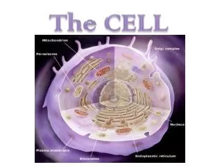
1 / 39

The Cell

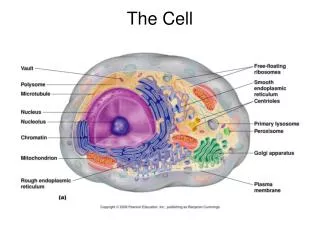
The Cell. Observed by Light and Electron Microscopy. Overview of the Cellular Organelles. Light Microscopy – Hematoxylin & Eosin (H&E) Stain. Basophilic (blue) Nuclei Eosinophilic (red) cytoplasm. Colorized low mag TEM of a cell. Red = Mitos Blue = Ribos Green = RER.
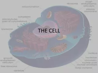
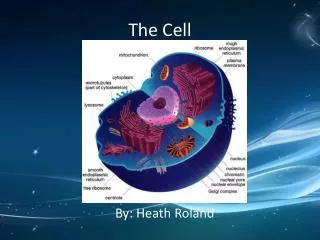
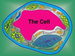
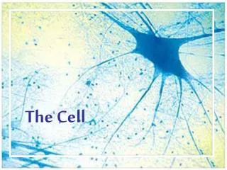
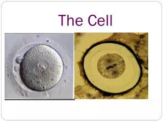
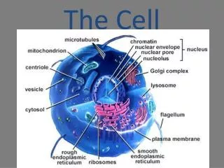
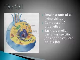
herman
Download Presentation

The Cell

An Image/Link below is provided (as is) to download presentation Download Policy: Content on the Website is provided to you AS IS for your information and personal use and may not be sold / licensed / shared on other websites without getting consent from its author. Content is provided to you AS IS for your information and personal use only. Download presentation by click this link. While downloading, if for some reason you are not able to download a presentation, the publisher may have deleted the file from their server. During download, if you can't get a presentation, the file might be deleted by the publisher.

E N D

Presentation Transcript


  1. The Cell Observed by Light and Electron Microscopy

  2. Overview of the Cellular Organelles

  3. Light Microscopy – Hematoxylin & Eosin (H&E) Stain Basophilic (blue) Nuclei Eosinophilic (red) cytoplasm

  4. Colorized low mag TEM of a cell Red = Mitos Blue = Ribos Green = RER

  5. Colorized TEM of a cell nucleus Arrow = Nucleolus Red = Mitos Green = RER

  6. Nuclear Structures

  7. Low mag TEM of a nuclei

  8. Nuclear pores (arrows) Nuclear pores(en face)

  9. Colorized TEM of nuclear pores (blue) en face

  10. High mag TEM of a nucleolus Pars fibrosa/granulosa (dense black) and fibrillar center (arrow)

  11. TEM of interphase nuclei and a mitotic cell with condensed chromosomes (arrow)

  12. Cytoplasmic Structures Ribosomes, RER and Glogi

  13. Ribosomes by light microscopy = basophilic cytoplasm

  14. TEMs of free ribosomes – 25 nm granules – in clusters called polysomes

  15. Colorized TEM of polysomes (yellow highlighted)

  16. Low mag TEM of rough endoplasmic reticulum (RER)

  17. Colorized higher mag TEM of RER (green)

  18. TEM of RER – arrows indicate cisternae

  19. Low mag TEM of RER and Golgi (Go)

  20. Colorized TEM of a Golgi system

  21. TEM of a Golgi system

  22. TEM of lysosomes (arrows) & a residual body Residual Body

  23. Cytoplasmic inclusions by Light Microscopy Basophilic granules (top picture) Eosinophilic granules (bottom picture)

  24. Mitochondria

  25. Colorized TEM of Mitochrondria (red)

  26. TEMs of Mitochrondrial shapes

  27. TEM of a Mitochrondrion Arrows = cristae

  28. The Cytoskeleton

  29. Light microscopy of mitotic spindles = Microtubules

  30. Colorized TEM of Cilia in cross section showing microtubules

  31. Immunofluorescence of Microtubules in cultured cells (green)

  32. TEM of Microfilaments (arrows)

  33. Immunofluorescence of microfilament stress fibers in cultured cells

  34. Cell Membrane

  35. High mag TEM of two cell membranes (unit membrane)

  36. Colorized TEM of two cell membranes (unit membrane)

  37. TEMs of membrane specializations Top 4 = microvilli Middle = Stereo cilia Bottom = Cilia

  38. TEM of Cilia (C) with microtubular cores forming from basal bodies (BB)

  39. High mag TEM of Cilia with microtubular cores (Fil) with the basal body (FiC) at top of image

More Related