Download
1 / 27
280 likes | 825 Views
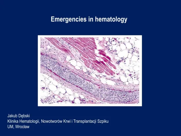
Emergencies in hematology. Jakub Dębski Klinika Hematologii, Nowotworów Krwi i Transplantacji Szpiku UM, Wrocław. Patient 1.

E N D
Emergenciesinhematology Jakub DębskiKlinika Hematologii, Nowotworów Krwi i Transplantacji SzpikuUM, Wrocław
Patient 1 Interview: 59-year-old patient with chronic lymphocytic leukemia diagnosed in 2009 in stadium - Rai II, Binet B was transferred from the Internal Medicine Department to the Department of Hematology for the treatment of pneumonia and significant progression of the disease. The patient who was previously untreated hematologically, did not agree to the proposed chemotherapyduringseveralvisitsintheoutpatientclinics - the last visit on 8.01.2014. In March 2015, he was hospitalized intheSurgery Unit due to phlegmon of theleftthigh. Then we found:leukocytosis 800 G / l, osteolytic lesions of left thighand pelvic bones , and in the performed on 17.03.2015 ultrasound examination of the abdominal cavity - hepatosplenomegaly with significantly enlarged lymph nodes, the largest with dimensions of 85 x 24 mm. The patient was advised to report urgently to the Hematology Clinic, but he did not. For about 3-4 days he hadbeenobservingcough, weakness, fever up to 40 C. Hospitalized intheInternalMedicine Unit between 29-30.04.2015 , in whichhe has been diagnosed with right-sided pneumonia with severe course and progression of untreated chronic lymphocytic leukemia.
Patient 1 At the reception on 30.04.2015:very severe condition, ECOG-3/4, drowsy, in a simple logical contact, conscious, aware, hereported a worsening of vision.In the physical examination:cachectic, right-sided rattling and crackling, left-sided crackles, RHA, tachypnoe 22 /min, abdomen arched above the chest level, soft, tender, without peritoneal symptoms, hepatosplenomegaly, numerous lymph node packages palpable through the abdominal wall, lymph nodes - cervical, axillary, inguinal – enlargedup to 2-3 cm in diameter, organized in packets, numerous scars and skin cavities of the left lower legdue to thephlegmon (cured).
Patient 1 Peripheralbloodmorphology: WBC 982.77 10*3/uL [4-10] , IG% 0.3 % , NEUT% 1.6 % [40-75] , LYMPH% 89.4 % [25-45] , MONO% 9 % [2-12] , EO% 0 % [1-6] , BASO% 0 % , NRBC% 0 % , IG# 2.72 10*3/uL , NEUT# 14.99 10*3/uL [1.6-7.5] , LYMPH# 878.61 10*3/uL [1-4] , MONO# 88.9 10*3/uL [0.08-1.2] , EO# 0.07 10*3/uL [0.04-0.6] , BASO# 0.2 10*3/uL [0-0.1] , NRBC# 0.41 10*3/uL , RBC 2.06 10*6/uL [4.5-5.9] , HGB 5.8 g/dL [14-18] , HCT 19.1 % [37-53] , MCV 92.7 fL [81-98] , MCH 28.2 pg [26-34] , MCHC 30.4 g/dL [31-37] , RDW-SD 63.2 fL [37-54] , RDW-CV % 18.9 % [11-16] , PLT 51 10*3/uL [130-400] , PDW 12.4 fL [9-17] , MPV 10 fL [9-13] , P-LCR 28.2 % [13-43] .
Patient 1 In laboratorytests:In peripheralbloodmorphology –Leukocytes 982.77 G / l,Hemoglobin 5.8 g / dl,Thrombocytes 51 G / l,?Procalcitonin 36.02 ng / ml,SaO2 82-84%,Biochemistry – Increasedlevel of glucose, CRP, LDH, urea, uricacid, ASPAT, ALP, total and direct bilirubin, magnesium, fibrinogen, D-dimersDecreasedlevel of potassium, total protein, albumin What should be the further managementof the patient?
Patient 1 The course of treatment:The patient was monitored, hydrated, and an urgent leukapheresis was performed followed by the first cycle of CVP (cyclophosphamide, vinblastine) chemotherapy in fractionated doses along with intravenous corticosteroids. RBC concentrate was transfused. In addition, broad-spectrum antibiotics and supportive care were used, achieving in the next days of stay an improvement in the general condition of the patient, along with moderate cytoreduction and a significantdecrease in inflammation parameters. Due to the rebound of leukocytosis on 8.05.2015, another leukapheresis procedure was performed and the second and third CVP chemotherapy courses were implemented on 8.05.15 and 28.05.15, followed by further cytoreduction and moderate reduction of the size of the lymph nodes, liver and spleen. Because of insufficient response achieved after the 3 cycles of CVP, the therapeutic protocol was changed to FC-R (fludarabine, cyclophosphamide, rituximab), the first cycle of which was introduced with good tolerance.
Patient 1 Leukapheresis - a method of selectivemechanicalpurification of plasmafrommorphoticelements - leukocytes (granulocytes, monocytes, leukemicblasts, CD34 + peripheralbloodstemcells) using a separator.
Hyperleukocytosis Definition: leukocytes > 100 G / lItleads to complications: LS - leukostasissyndrome DIC – disseminatedintravascularcoagulation TLS - tumor lysis syndromeTherisk of complicationsdepends on thebiology of leukemiccells. Epidemiology: - AML - acutemyeloid leukemia (10-20%) - subtypes M4, M5, microgranularvariant M3 - ALL - acutelymphoblastic leukemia (10-30%) - T-cellsubtypes, males, infants, 10-20 years of age (rarelyleukostasissyndrome) - BL - lymphoma / Burkitt's leukemia - mainly TLS- CLL - chroniclymphocytic leukemia – symptomatic > 400 G / l (rarelyleucostaticsyndrome) - CML - chronicmyeloid leukemia - mainlyintheblastcrisisphase
Leukostasissyndrome - symptomatic hyperleukocytosis (clinical concept, not laboratory) - disturbances of blood flow and tissue perfusion as a result of accumulation of leukemic cells in the lumen of microcirculation vessels, often with activation of coagulation- most often affects the lungs (30%) and CNS (40%) Symptoms: - dyspnoea, hypoxia, diffusevesicularhemorrhage, acute respiratory failure (ARDS)- visualimpairment, opticnerveedema, retinalhemorrhages- disturbances of consciousness, dizziness, tinnitus, drowsiness, headache, delirium, coma, focalneurologicaldeficits, intracranialhemorrhage- myocardial ischemia / ACS- acute limb ischemia- renalvein thrombosis, exacerbation of preexistingrenalfailure- priapism- intestinalinfarction- fever (due to cytokinereleaseor associated infection) - progression of respiratory distresssometimesinpatientsafterthe start of chemotherapy, (acute lysis pneumopathy)
Leukostasissyndrome In laboratorytests:pseudohypoxemiapseudohypoglycemiapseudohyperkalemiahypocalcaemiahyperphosphatemiahyperuricaemialacticacidosisDIC features - about 40% TLS features - about 10%
Leukostasissyndrome Treatment: - urgentleukapheresis – 2 procedures with an interval of 12-24 hours Indications for a leukapheresis:
Leukostasissyndrome Treatment:- cytoreduction: hydroxycarbamide 50-100 mg / kg, usually 2-4 g / day, reductioninthevalue of leukocytosis by 50-80% within 24-48 hours- chemotherapyinducingremission - implementedwhenleukocytes < 50 G / l - adequatehydration and prophylaxis of tumor lysis syndrome- management of thecoexist. intravascularcoagulationsyndrome- RBC concentratetransfusions - cautiously, slowlyduringoraftertheleukapheresisprocedure- Plateletconcentratetransfusions - maintenance of PLT level > 20-30 G / l- antibiotictherapy- glucocorticoids
Leukostasissyndrome Contraindications to leukapheresis:- acutepromyelocytic leukemia- cardiorespiratoryinsufficiency- severecardiacdiseases- severecoagulationdisorders
Disseminatedintravascularcoagulation (DIC) - 10-15% ptswithgeneralizedtumors, 15% with leukemia - generalizedactivation of coagulationwithfibrinolysisdisorders + coagulopathywithconsumption- intheclinicalpresentation: bleeding (60%) and thromboticcomplications - 3 types of DIC accompanyingtumors: procoagulant, hyperfibrinolytic, subclinical- diagnosis - serial analysis of plateletcounts, PT and aPTTcoagulationtimes, fibrindegradationmarkers: fibrinmonomers, FDP, D-dimers, fibrinogenconcentration
Disseminatedintravascularcoagulation (DIC) Management: - treatment of theunderlyingdisease- transfusions of blood products: RBC concentrate - whenthereis a significantloss of bloodFFP 10-15 ml / kg - activebleeding and elongation > 1.5-fold aPTTor PTFFP 15 ml / kg every 12-24 h orcryoprecipitate 1 unit / 10 kg every 24 h - activebleeding and fibrinogen < 1.0 g / l Plateletconcentrate 1-2 U / kg - PLT < 20 G / l or PLT < 50 G / l + hemorrhagicdiathesis- heparin (controversial) - should be consideredincaseswithpredominantthromboticsymptoms- fibrinolysisinhibitors - tranexamicacid 10-15 mg / kg in DIC withenhancedfibrinolysis- inacutepromyelocytic leukemia - DIC withhyperfibrinolysis, life-threateninghemorrhagiccomplicationsinabout 5% (65% intracranialbleeding / 32% pulmonarybleeding) – requiredapplication of ATRA (all-transretinoicacid) or ATO (arsenictrioxide)
Tumor lysis syndrome (TLS) - life-threateningmetabolicsyndromeresultingfromtherapidbreakdown of cancercells- high proliferativeactivity and high chemosensitivity of the tumor- spontaneous form - Burkitt'slymphoma, acute leukemia with high hyperleukocytosis- induced form - acute leukemia (ALL > 100 G / l, AML > 50 G / l), Burkitt'slymphoma, lymphoblasticlymphoma - renalfailureincreasestherisk of TLS - metabolicdisorders - hyperkalemia, hyperphosphatemia, hyperuricemia, hypocalcaemia, metabolicacidosis - symptoms - acuterenalfailure, arrhythmias / suddencardiacdeath , hypotension, heartfailure, convulsions, neuromuscularhyperactivity
Tumor lysis syndrome (TLS) Management:1. prophylaxis - hydration 3 l / m2 / 24 h 0.9% NaCl + allopurinol 600-900 mg / 24 h + eventuallyleukapheresis 2. treatment - diuresisforced by a loopdiuretic > 3 l / 24 h, hourly 100 ml / h + allopurinol 500 mg / m2 / day- inthecase of severehyperuricaemia - rasburicase (recombinantxanthineoxidase) 0.2 mg / kg / dayi.v. - hemodialysisinthe development of acuterenalfailure- correction of electrolytedisturbances
Hypercalcemia - increasedtotalcalciumconcentrations > 2.6 mmol / l (10.5 mg / dl) orionizedcalcium > 1.25 mmol / l (5 mg / dl) - inthecase of protein disorders (hypoproteinaemia, hypoalbuminaemia): calculationcorrectedcalciumconcentration (mg / dl) = [measuredcalciumconcentration (mg / dl) - albumin concentration (g / dl)] + 4 - inhyperproteinemia - possiblespurioushypercalcemia The most commoncausesare: - increasedosteolysisinplasmacellmyeloma- lymphomas and leukemia - paraneoplasticsyndromeorsevereosteolysis (PTH-independent) orproduction of 1,25(OH)2D3- bonetissueneoplasms, bonemetastases- primaryhyperparathyroidism- overdose of vitamin D3- persistentimmobilization- thiazidediuretics, theophylline- hyperthyroidism- adynamicbonediseaseindialysispatients
Hypercalcemia Mildhypercalcemia < 3.0 mmol / l (12 mg / dl) - asymptomaticModerate and severehypercalcemia 3.0 - 3.75 mmol / l (12-15 mg / dl) and rapidlyincreasing - hypercalcemicsyndrome:1. kidneyproblems - polyuria, hypercalciuria (nephrolithiasis, nephrocalcinosis)2. gastrointestinalsymptoms - lack of appetite, nausea, vomiting, constipation, gastric / duodenalulcer, pancreatitis, cholelithiasis3. cardiovascularsymptoms - arterialhypertension, tachycardia, arrhythmia, hypersensitivity to digitalis glycosides4. neuromuscularsymptoms - muscleweakness, weaktendonreflexes, transientfacialmuscleparalysis5. CNS symptoms - headache, depression, orientationdisorders, drowsiness, coma6. dehydrationIn a veryseverehypercalcemia > 3.75 mmol / l (15 mg / dl) - hypercalcemiccrisis: disturbances of consciousness, nausea, vomiting, abdominalpain, arrhythmias, dehydration, death. ECG - prolongation of the PQ interval, QT shortening, wide T wave,sometimestheOsbornwave
Hypercalcemia Management:- abundanthydration - 3-5 liters / day (~ 3 l / m2 / day) 0.9% NaCl- forceddiuresiswithfurosemide 20-40 mg iv, diuresis: 150-200 ml / h - reducingthecalciumreleasefrombones - calcitonin 100 UI 2-4 times a day iv orbisphosphonates (eg. pamidronate 60-90 mg / 2 h iv orzoledronate 4 mg / 15 min iv) ordenosumab- inhibition of calciumabsorptionfromthegastrointestinaltract - glucocorticosteroids (eg. hydrocortisone 100 mg iv every 6 h)- inseverecases – hemodialysisIn chronic tumor hypercalcemiainaddition:- mitramycin 25 ug / kg for 4-6 h i.v. or- galliumnitrate 100-200 mg / m2 for 24 hours for 4-5 days
Superior venacavasyndrome (SVCS) - syndrome of symptoms caused by impeding blood flow from the superior vena cava to the right atrium of the heart- 2-4% of patients with lung cancer and 2-4% with lymphoma The most frequent causes:- narrowing of the superior vena cava1.external pressure + tumor infiltration of the vein wall - lung cancer (60-80%), lymphoma (10-15%), metastasis of breast cancer, mediastinal germ cell tumors, thymomas 2. non-neoplastic causes - aortic aneurysm, chronic mediastinitis- superior venacavathrombosis - a catheter or secondary mediastinal tumor- right atrial tumor (rare)
Superior venacavasyndrome (SVCS) Symptoms:- swelling, erythema or bruising of the face- conjunctival hyperemia - swelling of the upper limbs - symptoms associated with increased intracranial pressure: headache and dizziness, visual disturbances, convulsions - permanent distentionof the jugular veins and positive Pemberton’s sign- dyspnea- hoarseness- difficulty in swallowing- stridorThe chest CT scan is the test of choice to diagnose SVCS.Chest X-ray is normal in 5-15% of cases.
Superior venacavasyndrome (SVCS) Symptomatic treatment:- dexamethasone 16-32 mg / d i.v. for 7 days, then dose reduction- control of dyspnea - morphine, midazolam, oxygen therapy- loop diuretic (eg. furosemide)- low molecular weight heparin - prophylactic or curativeCausative treatment:- urgent irradiation of the mediastinum - in the majority of cancers, the treatment of choice - chemotherapy - for chemosensitive cancers: lymphomas, lung cancer, germ cell tumors - introduction of the stent into the superior vena cava (removal of symptoms in> 75% of patients within 48 -72 h after surgery)- in the case of thrombosis - also consider thrombolysis
Differentiationsyndromeinacutepromyelocytic leukemia - life-threatening complication of the induction of acute myeloid leukemia M3 (promyelocytic) after application of ATRA or ATO- occurs inup to 25% of patients within 7-12 days of starting treatment- pathomechanism is not known, probably cytokine-related- symptoms: fever, increasing dyspnea, peripheral edema, skin rash, lung infiltration, fluid in the pleural cavity and pericardium, hypotension, renal insufficiency- risk increased by leukocytosis> 50 G / l - treatment: dexamethasone 2 x 10 mg iv until the symptoms disappear, sometimes dialysis is necessary and respiratory therapy- in patients with severe organ-related complications - temporary withdrawal of ATRA or ATO- mortality is around 1%
Neutropenicfever • neutropenia is the most common complication of anticancer treatment, it results from the myelotoxic action of chemo-radiotherapy or bone marrow infiltration by tumor cells- neutropenic fever - 10-50% of patients with solid tumors,> 80% with hematological malignanciesDefinition of neutropenicfever (febrileneutropenia): 1. oral temperature ≥ 38.3 C in single measurement or ≥ 38 C persistent for 1 hour 2. neutrophil count <500 / ul or predicted decrease to <500 / ul in 48 hoursPatients with NF rarely have local infection or they are weakly expressed. Most often, bacterial infections (Gram +> Gram-), less often fungal and viral.Blood-borne infection - 13-60% of patients, mortality 12-42%. The most serious forms of infection are: pneumonia, neutropenicenteritis, septic shock.
Neutropenicfever Management:- broad-spectrum, wide-spectrum empiric antibiotic therapy, in patients at high risk of complications and death: carbapenem or piperacillin with tazobactam or ceftazidime or cefepime iv - addition of antifungal medication (eg.fluconazole iv)- microbiological tests - identification of the pathogen responsible for infection is successful in 20-30% of patients- use of granulocyte growth factors G-CSF or GM-CSF, if the patient did not receive them as a primary or secondary prophylaxisRegular assessment of the patient's condition, effectiveness of antibiotic therapy, development of complications every 24-48 hoursismandatory.In case of improvement or identification of an infectious agent - de-escalation of antibiotic therapy.
Prophylaxis in afebrile patients with neutropenia 1) a sanitaryregime, hand and coughhygiene, sometimespatientisolation2) prophylacticuse of fluoroquinolone (ciprofloxacin, levofloxacin) - onlyinhigh-riskpatients3) prophylacticuse of antifungaldrugs (azoles, echinocandins) and antivirals (aciclovir) - onlyinpatientsundergoingallo-HCT and inducingremissionchemotherapyinacutemyeloid leukemia4) prophylacticuse of cotrimoxazoleinpatientswithriskfactors for Pneumocystisjiroveciinfection - purineanalogs, corticotherapy > 1 month5) prophylaxiswithG-CSF (filgrastim, lenograstim ) orGM-CSF (molgramostim, regramostim, sargramostim): - primary - recommendedalreadyduringthe first cycle of chemotherapy, whentherisk of developing febrileneutropenia > 20%, to consideralsointhecase of intensivechemotherapy - secondary - recommendedinpatientswithcomplications associated withneutropeniathatoccurredafterthe first chemotherapycycle, and reducingthedoses of cytotoxicdrugsordelayingtheiradministrationmayaffectoverallsurvival6) avoidance of prolongedenvironmentalcontact a high concentration of fungalmoldspores (constructions, old and neglectedbuildings)