Nervous System
430 likes | 560 Views
Explore the complexities of the nervous system, from sensory input to motor output. Learn about the central and peripheral nervous systems, neuron characteristics, and neurophysiology. Dive into the world of neurons, neuroglia, and supporting cells. Discover the role of myelination in nerve conduction and the impact of diseases like multiple sclerosis. Gain insights into neuronal structure and classifications. Unravel the essential functions like sensory, motor, and interneurons. Delve into the fascinating world of neurophysiology and the intricate workings of the nervous system.

Nervous System
E N D
Presentation Transcript
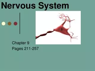
Nervous System Chapter 9 Pages 211-257
Chapter 9 Wordbytes • af- = toward 11.encephalo- = brain • arachn- = spider 12.gangli- = swelling • astro- = star 13. -glia = glue • auto- = self 14.mening- =membrane • dendro- = tree 15. micro- = small • di- = 2, through16.neuro- = nerve • ef- = away from17. –oid = similar to • enter- = intestines 18.oligo- = few • epen- = above 19. peri- = around • -ferrent = carried20.somat- = body
Nervous System Overview • Master controller and communicator for the body • Responsible for all behavior • 3 functions: • Sensory input monitors changes inside/outside of body • Integration processes and interprets, then decides what should be done • Motor output causes a response in effector organs
Organization—2 main parts: • Central Nervous System (CNS) = brain and spinal cord • Interprets incoming sensory info. and dictates motor responses • Peripheral Nervous System (PNS) = nerves from brain & in spinal cord • INPUT-Afferent or Sensory division • OUTPUT- Efferent or Motor division • Subdivided: Somatic (SNS—from CNS to skeletal muscles=voluntary) & Autonomic (ANS—regulate smooth & cardiac muscle, glands=involuntary)
Histology • Highly cellular—densely packed & tightly intertwined • 2 types of cells: • Neuron= nerve cell • Specialized for signal carrying & information processing • Neuroglia cells support, nourish & protect neurons • Neuroglia critical for homeostasis of interstitial fluid around neurons
Supporting cells (Neuroglia) • ~ half the volume of CNS • Cells smaller than neurons • Can multiply and divide and fill in brain areas • Do not conduct nerve impulses
Supporting Cells in CNS • Astrocytes most abundant and most versatile; blood-brain barrier • Oligodendrocytes (O lig o dendrocytes)have fewer branches; produce insulating myelin sheath in CNS • Microglia ovoid cells with thorny processes; provide defense (because immunity cells not allowed in CNS) • Ependymal cellssquamous/columnar cells with cilia; produce cerebrospinal fluid (CSF)
Supporting Cells in PNS • Schwann cells PNS cell support; produce & maintain myelin sheath, regenerate PNS axons • Satellite cells in PNS ganglia; support neurons in ganglia, regulate exchange of materials between neurons and interstitial fluid
Neuron Characteristics • They conduct nerve impulses from one part of the body to another • They have extreme longevity live/function for a lifetime • They are amitotic (a mi totic) lose their ability to divide • They have a high metabolic rate = need O2 and glucose
Neuronal Structure • Cell body nucleus, cytoplasm with typical organelles; most within CNS = protected by cranial bones & vertebrae • Dendrites short, highly branched input structures emerging from cell body = high surface area to receive signals • Axon may be short or long, only one per neuron; conducts away from cell body toward another neuron or effector • Emerges at cone-shaped axon hillock • Axon terminals at end of axon with synaptic bulbs
Figure 9.3 (Neurilemma) = impulse direction Pg. 216
Myelination • Axons covered with a myelin sheath • Many layered lipid & protein creating insulations • Increases speed of nerve conduction. • Formed by: • Schwann cells in PNS • Oligodendrocytes (O lig o dendrocytes) in CNS • Nodes of Ranvier (Ron v a)= gaps in the myelin • Nodes are important for signal conduction • Some diseases destroy myelin multiple sclerosis & Tay-Sachs
Multiple Sclerosis • What is it? https://health.google.com/health/ref/Multiple+sclerosis
Gray and White Matter • White matter- primarily myelinated axons • Gray matter- nerve cell bodies, dendrites, unmyelinated axons, axon terminals & neuroglia • Spinal cord gray matter is centrally located
Classification of Neurons • Structural according to # of processes (Fig. 9.6): • Multipolar 3 or more; most common, especially in CNS • Bipolar 2 processes (an axon and a dendrite) that extend from opposite sides; found in special sense organs • Unipolar 1 process that divides like a T; found in ganglia in PNS
Functional according to the direction impulse travels (Fig. 9.7) • Sensory (afferent) neurons transmit impulses from sensory receptors toward or into the CNS; mostly unipolar, with cell bodies in ganglia outside CNS • Motor (efferent) neurons carry impulses away from CNS to muscles and glands; multipolar, usually with cell bodies in CNS • Interneurons (association neurons) between motor & sensory neurons; most in CNS; 99% of neurons in body; mostly multipolar
Neurophysiology • Neurons are highly irritable = responsive to stimuli • When stimulated, an electrical impulse (action potential) is conducted along its axon • Action potential underlies all functional activities of the nervous system
Action Potentials • Action potentials = nerve impulses • Require a membrane potential • electrical charge difference across cell membrane – like a battery • Ion Channels allow ions to move by diffusion = current • If no action potential then resting cell has resting membrane potential
Ion Channels • Allow specific ions to diffuse across membrane • Move from high concentration to low or toward area of opposite charge • Leakage channels • Gated channels- require trigger to open • Voltage- Gated channels respond to a change in membrane potential
Resting Membrane Potential • Leakage channels • Cytosol high in K+ & interstitial fluid high in Na+(sodium –potassium pumps) • Leakage lets K+ through easily and Na+ poorly • inside is negative relative to outside • actual value depends on the relative leakage channel numbers
Graded Potentials • Short-lived, local changes to membrane potential • Cause current flows that decrease with distance • Magnitude varies with strength of stimulus
Action Potential (AP) • Generated by neurons and muscle cells • Series of active events • Channels actively open & close • Some initial event is required to reach a voltage threshold (~ = - 55 mv) • Stimulus = any event bringing membrane to threshold
Action Potential • Resting state • voltage-gated channels closed • Depolarizing phase- • membrane potential rises and becomes positive • Repolarizing phase- • potential restored to resting value ( PNa, PK) • Undershoot • Potassium permeability continues
Active Events • Stimulus to reach threshold • Na+ channel opens=> • Na+ ions enter=> • positive potential=> • Causes K+ channel opening => • repolarization
All- or –None Phenomenon • This sequence is always the same • If threshold then the same size of changes occur no larger or smaller APs • Stimulus must reach threshold to start • After one AP there is a short period before next can be triggered= absoluterefractory period each AP is a separate, all-or-none event; enforces one-way transmission of AP
Conduction of Nerve Impulses • Each section triggers next locally • Refractory period keeps it going the right direction • unmyelinated fiber- continuous conduction • With myelin- saltatory conduction • Can only be triggered at nodes of Ranvier • Myelinated fibers faster & larger neurons faster
The Syanpse • Synapse (to clasp or join)- junction that mediates information transfer from 1 neuron to another or from a neuron to an effector cell • Axodendritic or axosomaticsynapses – most synapses occur between the axonal ending of a neuron and the dendrites or cell body of other neurons
Synaptic Transmission – Electrical synapse • Sequence of events at synapse • Triggered by voltage change of the Action Potential • Sending neuron = presynaptic • Receiving neuron = postsynaptic • Space between = synaptic cleft • Neurotransmitter carries signal across cleft
Events at Synapse – Chemical synapse • AP arrives at presynaptic end bulb=> • Opens voltage gated Ca2+ channels=> • Ca2+ flows into cell • increased Ca2+ concentration => • exocytosis of synaptic vesicles=> • Neurotransmitter released into cleft • Diffuse across and bind to receptors in postsynaptic cell membrane
Synaptic Transmission • Binding at receptors • Chemical trigger of ion channels • May depolarize or hyperpolarize postsynaptic cell membrane • If threshold reached at axon hillock then postsynaptic cell action potential results
Synaptic Transmission • Finally the neurotransmitter must be removed from the cleft- • Diffusion away • Destroyed by enzymes in cleft • Transport back into presynaptic cell • Neuroglia destruction
Neurotransmitters • AcetylCholine (Ach)- common in PNS • Biogenic amines - Norepinephrine (NE), Dopamine (DA), serotonin, Histamine • Amino Acids- • Glutamate, Aspartate, gamma aminobutyric acid (GABA), glycine • Neuropeptides – endorphins • Novel Messengers - ATP/ Nitric oxide (NO)/ Carbon monoxide (CO)
Development of Neurons • P. 422-424 • Neuroblasts • Growth cone • Programmed cell death
Web sites: • http://www.sciencecases.org/chin/chin.asp • http://www.pbs.org/wgbh/nova/sciencenow/3204/01.html • http://www.getbodysmart.com/ap/nervoussystem/menu/menu.html • http://www.bbc.co.uk/science/humanbody/body/interactives/3djigsaw_02/index.swf?startPosition=nervous • http://learn.genetics.utah.edu/units/addiction/reward/madneuron.cfm • http://www.gpc.edu/~bbrown/peril/neurons/level1.htm


