Bone Fractures (Breaks)
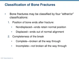
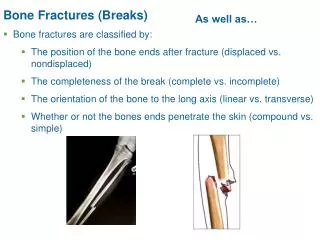
Bone Fractures (Breaks). As well as…. Bone fractures are classified by: The position of the bone ends after fracture (displaced vs. nondisplaced) The completeness of the break (complete vs. incomplete) The orientation of the bone to the long axis (linear vs. transverse)

Bone Fractures (Breaks)
E N D
Presentation Transcript
Bone Fractures (Breaks) As well as… • Bone fractures are classified by: • The position of the bone ends after fracture (displaced vs. nondisplaced) • The completeness of the break (complete vs. incomplete) • The orientation of the bone to the long axis (linear vs. transverse) • Whether or not the bones ends penetrate the skin (compound vs. simple)
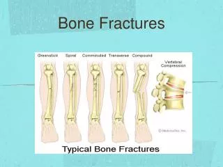
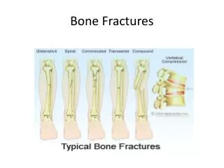
Common Types of Fractures Table 6.2.1
Common Types of Fractures Table 6.2.2
Common Types of Fractures Table 6.2.3
Stages in the Healing of a Bone Fracture 1. Hematoma formation (immediate – 1st day or 2) • Torn blood vessels hemorrhage • A mass of clotted blood (hematoma) forms at the fracture site • Site becomes swollen, painful, and inflamed Figure 6.13.1
Stages in the Healing of a Bone Fracture 2. Fibrocartilaginous callus forms (few days – week or 2) • Granulation tissue (soft callus) forms a few days after the fracture • Capillaries grow into the tissue and phagocytic cells begin cleaning debris Figure 6.13.2
Stages in the Healing of a Bone Fracture • Fibrocartilaginous callus forms when: • Osteoblasts and fibroblasts migrate to fracture • Fibroblasts secrete collagen fibers to connect bone (some become chondroblasts = cartilage) • Osteoblasts begin forming spongy bone • Osteoblasts furthest from capillaries secrete cartilaginous matrix that later calcifies
Stages in the Healing of a Bone Fracture 3. Bony callus formation (few weeks – 2 to 3 months) • More bone trabeculae appear in fibrocartilaginous callus • Fibrocartilaginous callus converts into a bony (hard) callus = firm union Figure 6.13.3
Stages in the Healing of a Bone Fracture 4. Bone remodeling (up to several months after bony callus) • Excess material on bone shaft exterior and in medullary canal removed • Compact bone laid down to reconstruct shaft walls Figure 6.13.4
Homeostatic Imbalances • Osteomalacia - inadequately mineralized bones (soft and weak) in adults • Main symptom = pain when weight is put on affected bone • Insufficient calcium in diet, or vitamin D deficiency • Rickets – inadequately mineralized bones of children • Bowed legs and deformities of the pelvis, skull, and rib cage • Insufficient calcium in the diet, or vitamin D deficiency • Example: Infants of breastfeeding mothers deficient in Vitamin D will also be Vitamin D deficient and develop rickets
Osteoporosis – bone-thinning disease (reabsorption outpaces deposition) • 30% of women over age 60-70 (70% by 80); 20% men over 70 • Makes bones fragile and fracture-prone (even a sneeze can cause fracture) • Often results in vertebral collapse (broken hips)
Osteoporosis: Treatment • Calcium and vitamin D supplements • Increased weight-bearing exercise • Hormone (estrogen) replacement therapy (HRT) slows bone loss • Increased risk of cancer and cardiovascular disease, yikes! • Statins increase bone mineral density
Paget’s Disease – haphazard and excessive bone deposit and resorption • High ratio of spongy to compact bone • Can cause spotty weakening of bone, pain, deformity (spine, pelvis, femur, skull) • Osteoclast activity wanes, but osteoblast activity continues • Possibly viral, 3% of Americans over 40 • Treated with drugs, calcitonin