Download
1 / 24
320 likes | 1.06k Views
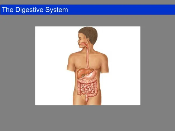
01.07.2015. Histology of Tongue, Liver & Pancreas. Dr. Archana Rani Associate Professor Department of Anatomy KGMU UP, Lucknow. Tongue. An accessory digestive organ. Composed of skeletal muscle covered with mucous membrane. Mucosa: Stratified squamous epithelium & lamina propria .

E N D
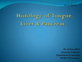
01.07.2015 Histology of Tongue, Liver & Pancreas Dr. Archana Rani Associate Professor Department of Anatomy KGMU UP, Lucknow
Tongue • An accessory digestive organ. • Composed of skeletal muscle covered with mucous membrane. • Mucosa: Stratified squamous epithelium & lamina propria. • Sulcusterminalis divides the dorsal aspect of tongue into anterior 2/3rd and posterior 1/3rd part.
Lingual Papilla • Lingual papilla: projections of lamina propria covered with stratified squamous epithelium. • May be keratinized. • Many papillae contain taste buds. • 4 types: Filiform, fungiform, circumvallate and foliate.
Types of Papillae • Filiform papilla: most numerous, conical with keratinized tips, no taste buds. • Fungiform papilla: mushroom shaped, highly vascularized connective tissue core, taste buds present. • Circumvallate papilla: surrounded by a circular trench, openings of the ducts of serous glands of Von Ebner. • Foliate papilla: not well developed in humans.
Taste Buds • Neurosensory epithelial structures embedded in the surface epithelium of fungiform and circumvallate papilla. • Appear as onion-like, oval, pale staining structures. • Extends through the full thickness of epithelium. • Opens on the surface through taste pore. • 3 types: Receptor cells, supporting cells and basal cells.
Muscles of the tongue • Contains striated muscle. • Muscle fibres are arranged in transverse, longitudinal and vertical bundles. • Loose connective tissue, adipose tissue and lingual glands are present in between the muscle bundles.
Liver • Modified exocrine gland. • Made up of liver cells (hepatocytes). • Hepatic lobules: hexagonal areas that form the structural & functional unit of liver. • Scanty connective tissue between lobules. • Each lobule is made up of cords of liver cells separated by sinusoids.
Liver • Portal canals contain branches of portal vein, hepatic artery & interlobular bile duct (portal triad). • Central vein: in the centre of each lobule, drains blood from lobules into hepatic veins.
Different types of liver lobules • A Classical liver lobule • B. A Portal liver lobule • C. A liver acinus
Liver • Glisson’s capsule: connective tissue covering of liver. • Bile canaliculi: spaces present between plasma membrane of adjacent liver cells. • Perisinusoidal space of Disse: separates the surface of liver cell from endothelial lining of the sinusoid.
Pancreas • Mixed gland i.e. consists of an exocrine & endocrine portion. • Exocrine part: secretes pancreatic juice. • Endocrine part: secretes hormones.
Histology of Exocrine Pancreas • Covered with a capsule. • Septas arise from capsule which divide the gland into many small lobules. • Interlobular connective tissue contains large ducts, blood vessels & nerve fibres. • Interlobular connective tissue surrounds the acini, small ducts & islets of Langerhans.
Pancreatic Acini • Serous in nature. • Lined by pyramidal cells and have small lumen. • Acinar cells: supranuclear region is filled with zymogen granules. Infranuclear region is intensely basophilic. • Extensive duct system: intralobular, interlobular & main duct. • Acinar lumen may show pale staining cells of intercalated duct (centroacinar cells).
Histology of Endocrine Pancreas • In the form of “islands”: Islets of Langerhans (lightly stained with H&E). • Scattered among the acini of exocrine part. • Cells of islets are arranged as anastomosing plates. • Islets contain 3 types of cells: Alpha, Beta & Delta which are seen with special stains.
References 1. diFiore’s Atlas of Histology with functional Correlations, 12th Edition. 2. Textbook of Human Histology. Inderbir Singh, 1st Edition. 3. Textbook of Histology. GP Pal, 3rd Edition.
MCQ Q1. The most numerous lingual papilla in humans are: a. Filiform b. Fungiform c. Circumvallate d. Foliate
MCQ Q2. Portal triad consist of all except: a. Branches of portal vein b. Branches of hepatic artery c. Central vein d. Bile duct
MCQ Q3. Space of Disse is present: a. Between sinusoidal epithelium and hepatocytes b. Between portal triad and hepatocytes c. Around central vein d. Between two adjacent plates of hepatocytes
MCQ Q4. Centroacinar cells are a characteristic feature of: a. Liver b. Pancreas c. Tongue d. Thyroid
MCQ Q5. Endocrine unit of pancreas is: a. Pancreatic acini b. Islets of Langerhans c. Von Ebner glands d. Centroacinar cells