Download
1 / 19
200 likes | 419 Views
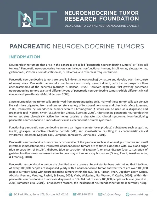
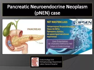
Pancreatic Neuroendocrine Neoplasm (pNEN) case. Case Presentation : Dr. Chatzellis Eleftherios MD Intern in Endocrinology. Endocrinology Unit Pathophysiology Department Laikon General Hospital. Case History - 2001. 58 year old male patient 2001 : abdominal pain

E N D
Pancreatic Neuroendocrine Neoplasm (pNEN) case • Case Presentation: • Dr. Chatzellis Eleftherios MDIntern in Endocrinology Endocrinology Unit Pathophysiology Department Laikon General Hospital
Case History - 2001 • 58 year old male patient • 2001: abdominal pain • CT scan:5cm tumor pancreatic tail Multiple focal lesions in the liver • FNAB (liver lesion) Well Differentiated NEN grade 2 (ki67 = 4%) • CgA = 5,2 nmol/l (<4), other markers negative • No secretory symptoms (non-functioning pNEN) • SRS (Octreoscan): avid uptake (Krenning score = 3) in both primary pNEN and liver metastases
Case History H&E stain ki67 Chromogranin A
Case History 2001-2005 • Sweden (Uppsala) Suggested surgery to reduce tumor burden and local complications • Patient refused • Chemotherapy (Streptozotocin + 5-FU) + Somatostatin analogues (SSA’s) • 1 year later (2002): DISEASE PROGRESSION (PD) • Addition of pegylated INF-a • Chemotherapy was discontinued after 2,5 years (mild renal impairment) • 2002-2005 : STABLE DISEASE (SD) • CgA = 10,5 nmol/l (<4)
Case History 2006-2008 • 2006: Hypercalcemia occurred for the first time • Ca = 11,8 mg/dl (8,5 - 10,1) • P = 1,98 mg/dl (2,5 - 4,5) • PTH = 2,54 pg/ml (10 - 65) • PTHrP = 84 pmol/l (<2) • CgA = 45 nmol/l (<4) • Imaging: DISEASE PROGRESSION (PD) • Increased SSA’s dosage [+peg-INFa] • Normalization Ca • 2006-2008: STABLE DISEASE (SD) - Biochemical control Humoral Malignancy-associated Hypercalcemia (PTHrP- related)
Case History 2008-2009 • 2008: peg-INFa discontinued (depression) • 2008: Hypercalcemia (12,9 mg/dl) + DISEASE PROGRESSION (PD) • 2008-2009: 177Lu-DOTATATE x5 cycles (25.6 GBq) • Increased SSA’s dosage • Addition of pasireotide 1200μg bid • Ca = 10,7 mg/dl, CgA=115 nmol/l, PTHrP=140 pmol/l • 2009: DISEASE PROGRESSION (PD) + biochemical relapse (Ca = 11,8 mg/dl)
Case History 2009-2010 • + Temozolomide + Capecitabine (CAP-TEM) • 2009-2010: STABLE DISEASE (SD) • Biochemical control of Ca required additional treatment: • SOM230 and SSA’s • Forced Diuresis • Prednisolone 40mg/d • Biphosphonates (i.v. zolendronate 4mg monthly) • Cinacalcet 90mg/d] • SIDE-EFFECTS: Proximal myopathy - muscle atrophy, patient immobilization (wheel chair), severe Diabetes Mellitus (~100 IU insulin/d)
Case History 2010 • Tumor burden reduction (cytoreduction) necessary • Biochemical control (Ca) • Reduce treatment side-effects • OPTIONS: • Surgery Patient still denied • PRRT Already performed (GFR, marrow toxicity) • RF Ablation Not applicable due to large liver lesions • Embolization (TAE/TACE) • Pre-embolization evaluation : Portal vein thrombosis • Selective approach (embolization of small branches of hepatic artery)
Case History - TAE Post-embolization Pre-embolization
TMZ + Capecitabine SSA’s Pasireotide 1200mcg/d Prednisolone 40mg/d Zolendronate 4mg/m Insulin treatment Ca = 10,2 mg/dl CgA = 50 nmol/l PTHrP = 38 pmol/l Post-embolization status Case History - TAE • TMZ + Capecitabine • SSA’s at ½ dosage • X • Prednisolone 8 mg/d • X • X
Case History 2010 • 6 months after TAE • Ca = 13,2 mg/dl • DISEASE PROGRESSION (PD) • Painful lump on left thigh T2 T1
Case History 2010 Bone scintigraphy Tc99m
Case History 2010 • BONE METASTASES vs BENIGN LESION • Bone Biopsy Brown tumor (in the context of prolonged PTHrP action on bone) • New liver lesion biopsy ki67 = 4%, IHC (+) for PTHrP H&E stain PTHrP Immunohistochemistry Liver metastases biopsy Bone biopsy
Case History 2010-2012 • Temozolomide (200mg/m2 Days 1-5 q4w) + Bevacizumab (10 mg/kg q2w) + Everolimus (10mg/d) • [+SSA’s] • Ca = 8,8 mg/dl • Temozolomide and Bevacizumab D/C after 6 months due to thrombocytopenia • 2010-2012: STABLE DISEASE (SD)
Case History 2012-2014 • 2012: DISEASE PROGRESSION (PD) • Ca=9,34 mg/dl, PTHrP = 50 pmol/l • Sunitinib 37,5 mg/d [+SSA’s] • 6 months later: Ca = 6,5 mg/dl !!! • 2012-2014: STABLE DISEASE (SD) • April 2014: Patient deceased † • (hepatic encephalopathy - malnutrition)
Pegylated INFa Pasireotide BEV-TEM STZ+ 5FU Everolimus Sunitinib PRRT CAP-TEM SSA’s PD PD PD PD PD PD Embolization • † Ca CgA PTHrP 2014 2012 2010 2012 2009 2009 2009 2006 2006 2001 2005 2008 2010 2010
Case synopsis • WD pNEN grade 2 Stage IV (liver metastases) • Change of functional status during disease course • Rare PTHrP secretion (paraneoplastic syndrome) • Even more rare: brown tumor due to PTHrP • Employment of several treatment agents/modalities to achieve both tumoral and biochemical control • Importance of cytoreductive interventions and novel molecular targeted therapies in controlling secretory symptoms/syndrome • Long survival despite metastatic disease at presentation (application of different therapeutic modalities)
Improving Survival of NET patients • James Yao, ENETS 2014