Download

1 / 131
1.43k likes | 2.09k Views
Myeloproliferative Neoplasm. (MPN). Guan Hongzai Department of Hematology E-mail: guanhongzai@qdu.edu.cn. INTRODUCTION.

E N D
Myeloproliferative Neoplasm (MPN) Guan Hongzai Department of Hematology E-mail: guanhongzai@qdu.edu.cn
INTRODUCTION The MPN are clonal haematopoietic stem cell disorders characterized by proliferation of one or more of the myeloid lineages (i.e. granulocytic, erythroid, megakaryocytic and mast cell) . Initially, MPN is characterized by hypercellularity of the BM with effective haematopoietic maturation and increased numbers of granulocytes, red blood cells and/or platelets in the PB.
INTRODUCTION Splenomegaly and hepatomegaly are common and caused by sequestration of excess blood cells or proliferation of abnormal haematopoietic cells. Despite an insidious onset each MPN has the potential to undergo a stepwise progression that terminates in marrow failure due to myelofibrosis, ineffectivehaematopoiesis or transformation to an acute blast phase.
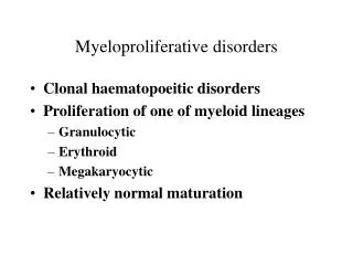
Common characteristics of MPD • Derive from the pathologic changes of multipotential stem cells • Accompanied with proliferation of one or more cell lineage(s). • Reciprocal transformation and concurrent with each other(1.ppt). • Extramedullary hematopoiesis and hepatosplenomegaly. • Cytomorphologic abnormalities in peripheral blood.
MPN include: Chronic myelogenous leukemia ( CML) Chronic neutrophilic leukaemia(CNL) Chronic eosinophilic leukaemia (CEL) PrimaryMyelofibrosis ( PMF ) Polycythemia vera ( PV ) Essential thrombocythemia ( ET ) Hypereosinophilic syndrome(HES) Mastocytosis Myeloproliferative neoplasm, unclassifiable(MPN-U)
PrimaryMyelofibrosis ( PMF ) Objective • Definition • Etiology and pathogenesis • Clinical features • Laboratory findings • Diagnosis and differential diagnosis
Definition Primary myelofibrosis ( PMF), also known as myelosclerosis (or agnogenic myeloid metaplasia), is a clonal myeloproliferative neoplasm (MPN) characterized by a proliferation of predominantly megakaryocytes and granulocytes in the bone marrow (BM) that in fully developed disease is associated with reactive deposition of fibrous connective tissue and with extramedullary haematopoiesis (EMH).
Definition There is a stepwise evolution from an initial prefibrotic phase characterized by a hypercellular BM with absent or minimal reticulin fibrosis to a fibrotic phase with marked reticulin or collagen fibrosis in the BM and often osteosclerosis. This fibrotic stage of PMF is characterized by leukoerythroblastosis in the blood with teardrop-shaped red cells, and by hepatomegaly and splenomegaly.
Etiology and pathogenesis The underlying cause is unknown in most PMF, the following agents may be reported: The JAK2V617F mutation may be found in 50% patients in the fibrotic phase. The stem cell change marrow hematopoietic disturbance increase of dysmorphic megakaryocytes in marrow release of cytokines ( PDGF, ECGF, TGF-β ) stimulate proliferation of fibroblast fibrous tissue accumulation.
Clinical Features • PMF usually occurs after age 40, the median age at diagnosis is 65 years. About ¼ of patients are asymptomatic at the time of diagnosis. • In symptomatic patients, fatigue, weakness, shortness of breath, palpitation, weight loss, night sweats, and bone pain are common presenting symptoms.
Clinical Features 3. Hepatomegaly is detectable in 50% of patients, splenomegaly is present in more than 90% and massive in one-third. 4. Severe anemia and hemorrhage are present in the advanced stage of the disease.
Laboratory Findings 1. Blood The peripheral findings that suggest a diagnosis of MF often include: • RBC • Normocytic-normochromic anemia is present in most patients. • Anisocytosis poikilocytosis, tear-drop red cells, basophilic stippling and nucleated red cell are consistently seen in the peripheral blood. • Reticulocytes usually range from 2% to 5%.
WBC • The total leukocyte count is usually normal or mildly increase, but may be as high as 100×109/L with neutrophilic granulocytosis. • Myelocytes and metamyelocytes are present in the blood of all patients, along with a low proportion of blasts ( 1%-5%).
WBC 3. Neutropilic alkaline phosphatase scores may be elevated in about two-thirds of the patients. 4. Basophils and eosinophils may be slightly increased. 5. Dysplastic leukocytes(Pelger-huët anomaly) may be present.
Thrombocytes • About one-third of patients have elevated platelet counts, and one-third have mild to moderate thrombocytopenia at the time of diagnosis. • Giant platelet, abnormal platelet granulation, and occasional circulating dwarf megakaryocytes are characteristic features of the disease. • ** About 10% of patients may present with pancytopenia. It is usually associated with intense marrow fibrosis.
Marrow Aspiration Marrow aspiration is usually unsuccessful because of fibrosis ( “dry tap”) Bone Marrow Biopsy Marrow biopsy is very important in the diagnosis of MF. It is often cellular and shows granulocytic, megakaryocytic, and erythron hyperplasia in the early stage. In intensely fibrotic marrow cellularity may be decreased almost replaced by fibrous tissues and collagen.
MF bone marrow biopsy Normal bone marrow
MF bone marrow biopsy AA bone marrow biopsy
Genetics and molecular findings • Chromosome abnormalities are evident in about 50% of the patients such as +8,-7,del(7q), del(11q), del(20q)and del(13q), but the ph chromosome is not present. • Approximately 50% of patients with PMF exhibit the JAK2V617F mutation.
Other Tests • Bleeding time can be prolonged. • Dyscontraction of clot. • Platelet adhesion and aggregation can be decreased. • Elevated serum levels of uric acid, lactic dehydrogenase(LDH) and alkaline phosphatase.
Diagnosis and Differential Diagnosis • Diagnosis • Splenomegaly. • Circulating immature myeloid cells and (or) nucleated red cells, accompanied with anisopoikilocytosis and tear-drop cell. • Bone marrow aspiration: dry tap or hypoplasia. • Extramedually hematopoiesis (spleen,liver,lymph nods) . • Bone marrow biopsy: diffuse fibrosis(essential condition )
Differential diagnosis Differential diagnosis between MF and CML MF CML WBC normal or mild increase >100 109/L RBC shape teardrop poikilocyte normal anisocytosis Nucleated RBC more seldom seen NAP score increase decrease(zero) Marrow smear dry tap (40%) myelocyte, meta- and stab granulocyte Marrow biopsy fibers, megakaryocyte myeloid hyperplasia Ph chromsome negative positive (95%) BCR/ABL gene negative positive
Polycythemia Vera ( PV ) Introduction PV is a clonal, chronic, progressive myeloproliferative disorder, often of insidious onset, characterized by an absolute increase in red cell mass and also usually by leukocytosis, thrombocytosis, and splenomegaly. The bone marrow is typically hypercellular and exhibits hyperplasia of myeloid, erythron, and megakaryocyte lineages.
Clinical features • PV usually has an insidious onset, most commonly at the age of 50 – 60 years. Presenting symptoms include dizziness, headache, eyes blurred, plethora, pruritus, weight loss, thrombosis, and gastrointestinal bleeding. • Hepatosplenomegaly present in 75% . One third of patients are hypertensive.
Laboratory Findings • Blood • RBC: male>6.5 1012/L; female >6.0 1012/L • Hb: male>180g/L; female >170g/L • Hct: male>0.54; female > 0.50 • The morphology of RBC are abnormal. Basophilic stippling and polychromatic cells increase in most patients
Laboratory Findings • WBC: (12-30) 109/L, with shift to left. NAP score raised (>100). • Platelet count increase in over 50 percent of patients, it is ranged from(400-500 ) 109/L.
Marrow • Bone marrow aspiration may be “dry tap”; The marrow shows deep red in color. • Hypercellularity or moderate hypercellularity with involvement of all lineages in most cases, marked in erythron, with normal shape. • Megakaryocytes increase. Clusters of five or more megakaryocytes may be seen.
Other Examinations • Red cell volume increase ( male>39ml/kg; female > 27ml/kg) • Blood viscosity increase (more than 5 — 6 times than that of normal.) • Vitamin 12 and uric acid usually raised. • Platelets have a characteristics functional defect. • Cytogenetic: clonal abnormalities in 20% of patients: dysploid, hyperdiploid, multiploid.
Diagnosis and differential diagnosis Diagnosis The most important diagnostic features are: 1. Splenomegaly and plethora. 2. RBC: male>6.5 1012/L; female >6.0 1012/L Hb: male>180g/L; female >170g/L 3. RBC volume: male>39ml/kg; female > 27ml/kg. 4. Hct: male > 0.54; female > 0.50 WBC >11 109/L, BPC > 400 109/L, NAP score>100. BM hypercellular with all lineages, marked in “E”. 5. Secondary erythrocytosis must be rule out. Diagnostic Criteria: 1+2+3+5 or 1+2+4+5
Differential diagnosis PV secondary erythrocytosis Hb and RBC Hct WBC N PLT N Splenomegaly + - BM proliferation E, G, Meg E NAP score N Serum VitB12 N EPO /N
Essential Thrombocythemia (ET) Introduction ET is a chronic myeloproliferative disorder characterized by a sustained proliferation of megakaryocytes, which leads to increased numbers of circulating platelets. In addition to platelet counts in excess, this disorder is characterized by profound marrow megakaryocyte hyperplasia, splenomegaly, and a clinical course punctuated by hemorrhagic or thrombotic episodes or both.
Clinical Features • Usually develops between ages 50 and 70. • Because platelet counts are now often done as a routine, the disorder is being discovered in patients who are asymptomatic. • Mild splenomegaly is found in 50-80 percent of patients. • Thrombotic complications: Arterial thrombosis occur more frequently than venous. The most common sites of arterial thrombosis involve the cerebrovascular, peripheral vascular, and coronary arterial circulations.
Clinical Features 5. Bleeding complications of ET are similar in nature to those seen in platelet or vascular disorders, occurring in superficial locations either spontaneously or after minimal trauma. The most common sites of bleeding are mucosal and gastrointestinal.
Laboratory Findings Blood 1. Platelet increase marked (more than 1000 109/L), MPV . 2. Platelet may be large, small, irregular, pale blue staining, hypogranular. Platelets occur always in clusters. 3. Mild leukocytosis mainly in mature granulocyte. NAP score increase. 4. Mild anemia are common.