Download
1 / 54
940 likes | 2.87k Views
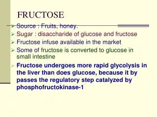
Fructose Metabolism. Catabolism of Fructose Considerable quantity of fructose is taken as sucrose (glucose-fructose), fruit juice and honey. This sugar is transformed into glucose or glycogen in liver and kidney and intestine Major pathway ,

E N D
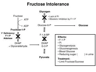
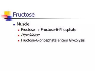
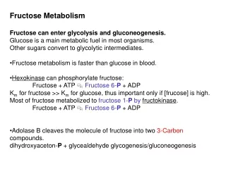
Catabolism of Fructose • Considerable quantity of fructose is taken as sucrose (glucose-fructose), fruit juice and honey. This sugar is transformed into glucose or glycogen in liver and kidney and intestine Major pathway, Fructokinase, in liver, muscle, kidney and intestine, has a high affinity for fructose than hexokinase giving fructose-1-phosphate. Minor pathway, Hexokinase has a low affinity to fructose and produces fructose-6-phosphate.
This sugar is present in milk containing diets and it is slowly transformed into glucose in liver. • It is synthesized from glucose in large quantities in actively lactating mammary gland and the blood and urine of lactating women may contain this sugar together with lactose. • Biochemical importance of galactose: • It enters in the formation of lactose. • It enters in the formation of glycolipids. • It enters in the formation of glycoproteins and proteoglycans
Normoglycemia: Fasting plasma glucose level (2 or more hours after last meal and up to 14 hours between meals, Post-Prandial) is 60-126 mg/dL measured by glucose oxidase In ideal individuals it should be less than the fasting level. Hypoglycemia:is a fasting blood glucose level below 45 mg/dL. Hyperglycemia:is a fasting blood glucose level above 126 mg/dL.
Sources and fates of blood glucose • Liver glycogen is enough to cover about 8 hours of fasting, whereas lactate from muscle glycogen can cover 25 hours. • About 40 % of the absorbed glucose is used for lipogenesis.
A. Tissue role in regulation of blood glucose Regulation of blood glucose
I. Gastrointestinal tract role: • It prevents hyperglycemia after carbohydrate meal by slowing the evacuation of the stomach. • Maximum rate of glucose absorption does not exceed 1 g/kg body weight/hour. Thus, absorption never exceeds the renal threshold • Upon contact with glucose the intestinal mucosa secretes certain factors into the blood, which stimulate insulin secretion.
II. Hepatic role: • It is the glucostat of the body. • After meal (during hyperglycemia), liver decreases blood glucose level by Glycogenesis, oxidation of glucose, non-essential amino acids synthesis and lipogenesis. • During fasting (during hypoglycemia), liver increases blood glucose by Glycogenolysis and gluconeogenesis.
III. Role of Muscles and adipose tissue: • After meal muscles prevent hyperglycemia by utilizing glucose in glycogenesis while adipose tissue utilizes blood glucose in lipogenesis. • On glycogenolysis, muscle supply lactate as a gluconeogenic substrate, whereas, during fasting adipose tissue supply glycerol as a gluconeogenic substrate
IV. Role of Kidneys: • It maintains the blood glucose by preventing its loss in the urine. • It adds little glucose to the blood by gluconeogenesis during fasting. • It lowers blood glucose if its level exceeds renal threshold (160 -180 mg/dl) by excretion in urine.
B. Hormonal regulation I. Insulin: • It secretion from β-cells of islets of Langerhans of pancreas is stimulated mainly by glucose. • Its secretion is inhibited by epinephrine. • It is inactivated in liver by GSH-insulin transhydrogenase and in liver, kidney and placenta by specific proteases. • It is the only hypoglycemic hormone.
A-On carbohydrate metabolism: • Increases glucose uptake by increasing glucose transporters in muscles and adipose tissue. • Stimulates glucose oxidation • Inhibits gluconeogenesis • Stimulates Glycogenesis • Inhibits Glycogenolysis
B-On lipid metabolism: • Insulin stimulates lipogenesis and ketolysis, whereas, it inhibits lipolysis. C-On protein metabolism: Increases amino acid uptake by the cells and stimulates protein synthesis. D-On nucleic acid metabolism: Insulin stimulates nucleic acid synthesis.
II. Glucagon: • Its secretion from α-cells of Langerhans is induced by hypoglycemia It cause hyperglycemia by: • Stimulation of Glycogenolysis: Glucagon has no effect on muscle phosphorylase (no glucagon receptors on muscles). • Stimulation of gluconeogenesis from pyruvate, lactate and amino acids by stimulating phosphoenol pyruvate carboxykinase.
III. Glucocorticoids ACTH stimulates their secretion by adrenal cortex. They are hyperglycemic by: • Stimulate protein breakdown in peripheral tissues into amino acids and stimulate transamination and gluconeogenesis key. • Decrease peripheral glucose uptake and utilization except in brain, intestine, RBCs, heart, liver and kidney. • Maintain liver glycogen, thus they are catabolic on peripheral tissues and anabolic on liver.
III. Glucocorticoids ACTH stimulates their secretion by adrenal cortex. They are hyperglycemic by: • Stimulate protein breakdown in peripheral tissues into amino acids and stimulate transamination and gluconeogenesis key • Decrease peripheral glucose uptake and utilization except in brain, intestine, RBCs, heart, liver and kidney. • Maintain liver glycogen, thus they are catabolic on peripheral tissues and anabolic on liver.
IV. Adrenaline and nor adrenaline Their secretion by adrenal medulla is stimulated by hypoglycemia and are hyperglycemic (more than 180 mg/dl in stress conditions, where blood sugar level is normal) by: Stimulation of liver glycogenolysis and glucose output into blood when blood glucose level falls below 60 mg/dl. Stimulation of muscle glycogenolysis releases large amount of lactate that is used for gluconeogenesis. They inhibit release of insulin from Beta-cells.
Hyperglycemia Variations in normal blood glucose
It is the rise of blood glucose level above 140 mg/dl. Causes: A-Deficiency of insulin in: • Diabetes mellitus (commonest cause). • Experimental or surgical pancreatectomy (removal of pancreas). • Alloxan and streptozotocin injection that destroy b-cells. • Pancreatitis and pancreatic cancer.
B-Increased anti-insulin hormones: • ACTH and glucocorticoids: as in adrenal cortical tumors and Cushing's syndrome. Also, other stresses, e.g., sepsis, some infectious diseases, anesthesia, asphyxia and convulsions. • Adrenaline: as in emotions and stress • TSH and thyroxine as in hyperthyroidism. • Pituitary growth hormone: as in Acromegaly.
C-Dietary or Alimentary, : high carbohydrate diet especially rich in simple sugars. D-Drug-induced, : e.g., chronic use of corticosteroids
Hypoglycemia It is the decrease of blood sugar level below 40 mg/dl. A-Glycogen storage diseases: Causes fasting hypoglycemia. B-Excess of insulin as in: • Overdose of insulin during treatment of diabetes mellitus lead to hypoglycemia. • Missing a meal during treatment with insulin. • Insulinoma, a tumor of Beta-cells.
C-Decrease of anti-insulin hormones in cases of: • Glucocorticoids as in Addison’s disease. • Pituitary hormones as in panhypopituitarism. • Thyroxin as in hypothyroidism. • Liver diseases sever exercises, alcoholism, tumors secreting IGFs and leucine sensitivity.
Effect of hypoglycemia: • Hypoglycemia is a very dangerous condition because glucose is the major fuel of the brain. • Hypoglycemia causes confusion and dizziness. • If blood glucose level is decreased below 40 mg/dl hypoglycemic coma will occur.
Presence of glucose in a detectable amount by qualitative Benedict's test is called Glucosuria. Normally, there is about 0.5 g of glucose in urine that is undetectable. Glucose starts to appear in urine when its blood level exceeds that maximum renal reabsorption limit, i.e., renal threshold that is 160 - 180 mg/dL. Reabsorption is an active Na+-dependent glucose transporter-mediate process. Presence of fructose in urine is called fructosuria, lactose is called lactosuria, and galactose is called galactosuria.
Causes of Glucosuria Diabetes mellitus Alimentary Glucosuria due to intake of high carbohydrate diet. Emotional Glucosuria: Surgical and experimental diabetes mellitus induced by: Total or subtotal pancreatectomy and alloxan or streptozotocin injection.
It is a chronic polygenic syndrome with impaired carbohydrate metabolism due to deficiency or ineffectiveness of insulin or decreased insulin/anti-insulin ratio leading to chronic hyperglycemia and Glucosuria along with secondary changes in metabolism of protein, lipids, water and electrolytes. It has grave consequences if not treated.
Causes: Defective processing of proinsulin into insulin, or peripheral resistance to insulin action due to defects in its receptor and sub-receptor mediators. Increased production of the anti-insulin hormones, as in Cushing's syndrome, acromegaly, increased glucagon and pheochromocytoma and stresses such as pregnancy and obesity. It has a genetic autosomal recessive predisposition. Autoimmunity, pancreatitis and pancreatic cancer. Viral infection, e.g., mumps and influenza. Diet overeating with under activity, particularly for carbohydrates.
Type 1 diabetes Type 1 diabetes mellitus is characterized by loss of the insulin-producing beta cells of the islets of Langerhans in the pancreas leading to insulin deficiency. This type of diabetes can be further classified as immune-mediated. The majority of type 1 diabetes is of the immune-mediated nature, where beta cell loss is a T-cell mediated autoimmune attack. Type 1 diabetes can affect children or adults but was traditionally termed "juvenile diabetes" because it represents a majority of the diabetes cases in children.
Type 2 diabetes Type 2 diabetes mellitus is characterized by insulin resistance which may be combined with relatively reduced insulin secretion. The defective responsiveness of body tissues to insulin is believed to involve the insulin receptor. However, the specific defects are not known. Type 2 diabetes is the most common type. In the early stage of type 2 diabetes, the predominant abnormality is reduced insulin sensitivity.
Gestational diabetes Gestational diabetes mellitus (GDM) resembles type 2 diabetes in several respects, involving a combination of relatively inadequate insulin secretion and responsiveness. It occurs in about 2%–5% of all pregnancies and may improve or disappear after delivery. Gestational diabetes is fully treatable but requires careful medical supervision throughout the pregnancy.
Signs and symptoms The classical symptoms of diabetes are polyuria (frequent urination), polydipsia (increased thirst) and polyphagia (increased hunger). Symptoms may develop rapidly (weeks or months) in type 1 diabetes while in type 2 diabetes they usually develop much more slowly and may be subtle or absent.
Prolonged high blood glucose can cause glucose absorption in the lens of the eye, which leads to changes in its shape, resulting in vision changes. Blurred vision is a common complaint leading to a diabetes diagnosis; type 1 should always be suspected in cases of rapid vision change, whereas with type 2 change is generally more gradual, but should still be suspected. A number of skin rashes can occur in diabetes that are collectively known as diabetic dermadromes.
Complications of diabetes mellitus All forms of diabetes increase the risk of long-term complications. The major long-term complications relate to damage to blood vessels. Diabetes doubles the risk of cardiovascular disease.The main "macrovascular" diseases (related to atherosclerosis of larger arteries) are ischemic heart disease (angina and myocardial infarction), stroke and peripheral vascular disease.
Diabetic retinopathy, which affects blood vessel formation in the retina of the eye, can lead to visual symptoms, reduced vision, and potentially blindness. Diabetic nephropathy, the impact of diabetes on the kidneys, can lead to scarring changes in the kidney tissue, loss of small or progressively larger amounts of protein in the urine, and eventually chronic kidney disease requiring dialysis.
Diabetic neuropathy is the impact of diabetes on the nervous system, most commonly causing numbness, tingling and pain in the feet and also increasing the risk of skin damage due to altered sensation. Together with vascular disease in the legs, neuropathy contributes to the risk of diabetes-related foot problems (such as diabetic foot ulcers) that can be difficult to treat and occasionally require amputation.
Diagnosis Diabetes mellitus is characterized by recurrent or persistent hyperglycemia, and is diagnosed by demonstrating any one of the following: • Fasting plasma glucose level ≥ 126 mg/dL. • Plasma glucose ≥ 200 mg/dL two hours after a 75 g oral glucose load as in a glucose tolerance test. • Symptoms of hyperglycemia and casual plasma glucose ≥ 200 mg/dL. • Glycated hemoglobin (HbA1C) ≥ 6.5%.
Diabetes in dogs and diabetes in cats • In animals, diabetes is most commonly encountered in dogs and cats (Middle-aged animals). • Female dogs are twice as likely to be affected as males, while according to some sources male cats are also more prone than females. • In both species, all breeds may be affected, but some small dog breeds are particularly likely to develop diabetes.
The symptoms may relate to fluid loss and polyuria. • Diabetic animals are more prone to infections. • The long-term complications recognised in humans are much rarer in animals. • The principles of treatment (weight loss, oral antidiabetics, subcutaneous insulin) and management of emergencies (e.g. ketoacidosis) are similar to those in humans.
A glucose tolerance test is a medical test in which glucose is given and blood samples taken afterward to determine how quickly it is cleared from the blood. The test is usually used to test for diabetes, insulin resistance, and sometimes reactive hypoglycemia and acromegaly, or rarer disorders of carbohydrate metabolism. In the most commonly performed version of the test, an oral glucose tolerance test (OGTT), a standard dose of glucose is ingested by mouth and blood levels are checked two hours later.
Many variations of the GTT have been devised over the years for various purposes, with different standard doses of glucose, different routes of administration, different intervals and durations of sampling, and various substances measured in addition to blood glucose.
Preparation The patient is instructed not to restrict carbohydrate intake in the days or weeks before the test. The test should not be done during an illness. Usually the OGTT is performed in the morning. The patient is instructed to fast (water is allowed) for 8–12 hours prior to the tests.