Download
1 / 16
160 likes | 267 Views
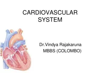
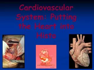
Cardiovascular System: Putting the Heart into Histo. Mesothelium. Lecture 9, Slide 1. 3 Layers Endocardium Myocardium Epicardium. L9, S2. Fibrous Trigone Forms the base of tissue from which the valves are anchored Separates atria from ventricles

E N D
Mesothelium Lecture 9, Slide 1
3 Layers Endocardium Myocardium Epicardium L9, S2
Fibrous Trigone Forms the base of tissue from which the valves are anchored Separates atria from ventricles Atria and ventricle only communicate through the AV bundle L9, S2
DFIACT Adipose Coronary vessels and cardiac nerves Externally lined by mesothelium Simple squamous cells Secrete serous fluid at visceral and parietal pericardium Epicardium L9, S3
M Z I A H L9, S5
Myocardium L9, S4 L9, S6
Inner layer Simple squamous epithelium and CT Elastic lamina Middle layer DCT with smooth muscle Subendocardial layer Purkinje fibers Continuous with myocardium Endocardium L9, S7 Masson’s Trichrome
Purkinje Fibers L9, S11 • Branches of the AV bundle of His • Cardiac muscle modified for conducting impulses • Located in the subendothelium
Hypertrophied cardiac muscle fibers • One or two nuclei centrally situated • Rich in glycogen and mitochondria L9, S12
Tunica Intima Internal Elastic Membrane Tunica Media External Elastic Membrane Tunica Adventitia 1 2 3 L9, S13
Tunica Intima Endothelium and BM Internal Elastic Membrane Tunica Media Reticular or Elastic fibers SMCs connecting elastic fibers External Elastic Membrane Tunica Adventitia Diffuse Vasa vasorum Nervi vascularus The Aorta: An Elastic Artery L9, S15 L9, S16
F • A = Aorta • B = Vein • C = Artery • D = Nerve • E = Brown adipose • F = White adipose • G = CT A B C G D E L9, S17
Aorta • Weigert Stain of the Elastic lamina L9, S19
Q1 E. None of the above A B D C L9, S4 L9, S6
Q2 and Q3 A B L9, S12