The Cardiovascular System: Blood Vessels
The Cardiovascular System: Blood Vessels Chapter 19 Introduction The blood vessels of the body form a closed delivery system that begins and ends at the heart


The Cardiovascular System: Blood Vessels
E N D
Presentation Transcript
The Cardiovascular System:Blood Vessels Chapter 19
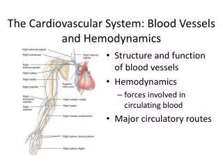
Introduction • The blood vessels of the body form a closed delivery system that begins and ends at the heart • Often compared to a plumbing system, it is a far more dynamic system of structures that pulse, constrict and relax and even proliferate to meet changing body needs
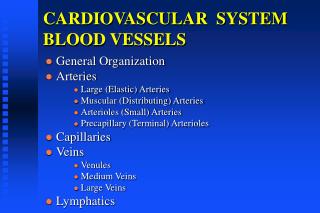
Blood Vessel Structure & Function • The major types of blood vessels are • Arteries • The large distributing vessels that bring blood to the body • Capillaries • The tiny vessels that distribute blood to the cells • Veins • The large collecting vessels that bring blood back to the heart • Intermediate vessels connect • Arterioles bring blood to the capillaries • Venules drain blood from the capillaries
Blood Vessel Structure & Function • The pattern of distribution starts with arteries to arterioles to capillaries to venules to veins • The blood vessels in the adult human body carry blood in a distribution network that is approximately 60,000 miles in length • Only capillaries come into intimate contact with tissue cells and serve cellular needs
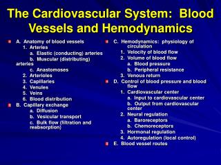
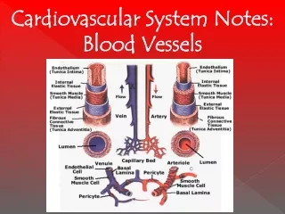
Blood Vessel Walls • The walls of blood vessels are composed of three distinct layers or tunics • The tunics surround a central opening called a lumen
Blood Vessel Walls • The innermost tunic is the tunica intima • This tunic contains the endothelium, the simple squamous endothelium that lines all vessels • Its flat cells fit closely together, forming a slick surface that minimizes friction as blood moves through the vessel lumen Tunica adventitia
Blood Vessel Walls • In blood vessels larger than 1 mm in diameter, a sub- endothelial layer of loose connective tissue, subendothelial layer, (basement membrane) supports the endothelium
Blood Vessel Walls • The middle tunic, the tunica media, is mostly circularly arranged smooth muscle cells and sheets of elastin • The activity of the smooth muscle is regulated by vasomotor nerve fibers of the sympathetic division of the autonomic nervous system Tunica media
Blood Vessel Walls • Depending on the needs of the body, the vasomotor fibers can cause vaso-constriction or vasodilation • The activities of the tunica media are critical in regulating circulatory dynamics • Generally, the tunica media is the bulkiest layer in arteries, which bear the chief responsibility for maintaining blood pressure and continuous blood circulation
Blood Vessel Walls • The outermost layer of a blood vessel is the tunica externa • This tunic is composed largely of loosely woven collagen fibers that protect blood vessels and anchor it to surrounding structures Tunica externa
Blood Vessel Walls • The tunica externa is infiltrated with nerve fibers and lymphatic vessels and, in larger vessels, a system of tiny blood vessels • These vessels, the vasa vasorum nourish the external tissues of the blood vessel wall Tunica externa
Arteries • Arteries are vessels that carry blood away from the heart • All arteries carry oxygen rich blood with the exception of those in the pulmonary circuit • Blood proceeds to the tissues through • Elastic arteries • Muscular arteries • Arterioles
Elastic (Conducting) Arteries • Elastic arteries are thick walled arteries near the heart - the aorta and its major branches • These arteries are the largest in diameter and the most elastic • A large lumen allows them to serve as low resistance pathways that conduct blood from the heart to medium-sized arteries and thus are called conducting arteries
Elastic (Conducting) Arteries • The elastic arteries contain more elastin than any other type of vessel • While present in all three layers, the tunica media contains the most • The abundant elastin enables these arteries to withstand and smooth out large pressure fluctuations by expanding when the heart forces blood into them and then recoiling to propel blood onward into the circulation when the heart relaxes
Elastic (Conducting) Arteries • Elastic arteries also contain substantial amounts of smooth muscle, but they are relatively inactive in vasoconstriction • Because elastic arteries expand and recoil passively to accommodate changes in blood volume, the blood is kept under pressure • Thus, blood flows continuously rather than starting and stopping with each heart beat
Muscular (Distributing) Arteries • The muscular distributing arteries deliver blood to specific body organs and account for most of the named arteries • Proportionately, they have the thickest media of all vessels • Their tunica media contains relatively more smooth muscle and less elastic tissue than that of elastic arteries • They are more active in vasoconstriction and are less distensible
Muscular (Distributing) Arteries • As in all vessels, concentric sheets of elastin occur within the tunica media of muscular arteries although these sheets are not as thick or abundant as those of elastic arteries
Muscular (Distributing) Arteries • A feature unique to muscular arteries, especially thick sheets of elastin lie on each side of the tunica media • An external elastic lamina lies between the tunica media and tunica externa
Muscular (Distributing) Arteries • The elastin in muscular arteries, like that in elastic arteries, helps dampen the pulsatile pressure produced by the heartbeat
Arterioles • Arterioles have a lumen diameter from 0.3 mm to 10 m, and are the smallest of the arteries • Larger arterioles exhibit all three tunics, but their tunica media is chiefly smooth muscle with a few scattered muscle fibers • The smaller arterioles that lead into capillary beds, are little more than a single layer of smooth muscle cells spiraling around the endothelial lining
Arterioles • The diameter of each arteriole is regulated in two ways: • Local factors in the tissues signal the smooth musculature to contract or relax, thus regulating the amount of blood sent downstream to each capillary bed • Sympathetic nervous system adjusts the diameter of arterioles throughout the body to regulate systemic blood pressure
Capillaries • The microscopic capillaries are the smallest blood vessels • In some cases, one endothelial cell forms the entire circum- ference of the capillary wall • The average length of a capillary is 1 mm and the average diameter is 8-10 m
Capillaries • Capillaries have a lumen just large enough for blood cells to slip through in single file
Capillaries • Capillaries are the body’s most important blood vessels because they renew and refresh the surrounding tissue fluid (interstitial fluid) with which all cells in the body are in contract • Capillaries deliver to interstitial fluid the oxygen and nutrients that cells need while removing carbon dioxide and nitrogenous wastes that cells deposit in the fluid
Capillaries • Given their location and the thinness of their walls capillaries are ideally suited for their role of providing access to nearly every cell • Along with the universal functions just described some capillaries also perform site-specific functions • Lungs: gas exchanges • Endocrine glands: pick up hormones • Small intestine: nutrients • Kidneys: removal of nitrogenous wastes
Capillary Beds • A capillary bed is a network of the body’s smallest vessels that run throughout almost all tissues, especially the loose connective tissue • This flow is also called a microcirculation
Capillary Beds • In most body regions, a capillary bed consists of two types of vessel a vascular shunt (meta- arteriole) and true capillaries
Capillary Beds • The terminal arteriole leads into a metarteriole which is directly continuous with the thorough- fare channel
Capillary Beds • The thoroughfare channel joins the post- capillary venule that drains the capillary bed
Capillary Beds • The true capillaries number 10 to 100 per capillary bed, depending on the organ served • Branch from metarteriole to thoroughfare channel
Capillary Beds • A cuff of smooth muscle fibers, called a pre- capillary sphincter surrounds the root of each capillary at the metarteriole and acts as a valve to regulate the flow of blood into the capillary
Capillary Beds • When the precapillary sphincters are relaxed, blood flows through the true capillaries and takes part in exchanges with tissue cells
Capillary Beds • When the precapillary sphincters are contracted, blood flows through the shunts and bypasses the tissue cells
Capillary Beds • Most tissues have a rich supply, but there are a few exceptions • Tendons and ligaments / poorly vascularized • Cartilage / from adjacent connective tissue • Epithelia / from adjacent connective tissue • Cornea / nourished by aqueous humor
Capillary Beds • The relative amount of blood entering a capillary bed is regulated by vasomotor nerve fibers and local chemical conditions • A capillary bed may be flooded with blood or almost completely bypassed, depending on conditions in the body or in that specific organ • Example of shunting blood from digestive organs to skeletal muscles
Capillary Permeability • The structure of capillaries is well suited for their function in the exchange of nutrients and wastes between the blood and the tissues through the tissue fluid • A capillary is a tube consisting of thin endothelial cells surrounded by a basal lamina • The endothelial cells are held together by tight junctions and occasional desmosomes
Capillary Permeability • Tight junctions block the passage of small molecules, but such junctions do not surround the whole perimeter of the endothelial cells • Instead, gaps of unjoined membrane called intercellular clefts occur through which small molecules exit and enter the capillary
Capillary Permeability • External to the endothelial cells, the delicate capillary is strengthened and stabilized by scattered pericytes
Capillary Permeability • The pericytes are spider shaped cells whose thin processes form a network that is widely spaced so as to not to interfere with capillary permeability
Capillary Permeability • Structurally there are three types of capillaries • Continuous • Fenestrated • Sinusoidal
Continuous Capillaries • Continuous capillaries are abundant in the CNS, skin and muscles and are the most common • They are continuous in the sense that their endothelial cells provide an uninterrupted lining
Continuous Capillaries • Adjacent cells are joined laterally by tight junctions • However, these are usually incomplete and leave gaps of unjoined membrane called intracellular clefts that are just large enough to allow limited passage of fluids
Fenestrated Capillaries • Fenestrated capillaries have fenestrations (pores) spanning the endothelial cells • Fenestrated capillaries occur only where there are exceptionally high rates of exchange of small molecules between blood and the surrounding tissue
Fenestrated Capillaries • The fenestrations are usually covered by a thin diaphragm but this variety has much greater permeability to fluids and small solutes • Fenestrated capillaries are found where active capillary absorption or filtrate formation occurs
Fenestrated Capillaries • Fenestrated capillaries are found in the small intestine to receive digested nutrients • These capillaries are also found in the synovial membranes of joints to allow water molecules to exit the blood to form synovial fluid Intercellular clefts
Routes of Capillary Permeability • Molecules pass into and out of capillaries via four routes • Direct diffusion through endothelial cell membranes • Through the intercellular clefts • Through cytoplasmic vesicles or caveolae • Through fenestrations in fenestrated capillaries
Routes of Capillary Permeability • Most exchange of small molecules is thought to occur through intercellular clefts • Caveolae apparently transport a few larger molecules, such as small proteins • Carbon dioxide and oxygen seem to be the only important molecules that diffuse directly through endothelial cells because these uncharged molecules easily diffuse through lipid containing membranes of cells
Low Permeability Capillaries • The blood-brain barrier prevents all but the most vital molecules(even leukocytes) from leaving the blood and entering brain tissue • The blood-brain barrier derives its structure from the capillaries of the brain • Brain capillaries have complete tight junctions, so intercellular clefts are absent
Low Permeability Capillaries • Brain capillaries are continuous, not fenestrated and they also lack caveolae • Vital capillaries that must cross brain capillaries are “ushered through” by highly selective transport mechanisms in the plasma membranes of the endothelial cells