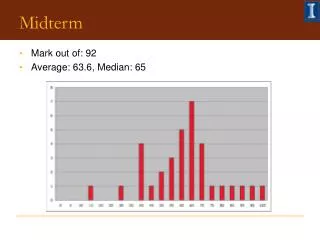
Midterm
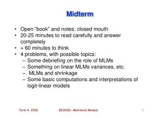
Midterm. Nov. 8 th Content, ch. 8, 9, 10, 11, 13, 14, 15. Muscle, Nervous Tissue, Blood, Circulatory system, endocrine system, integument, respiratory system ( not digestive system). We have talked about the mucosa in the fundic region. The gastric pits The fundic glands Mucus neck cells

Midterm
E N D
Presentation Transcript
Midterm • Nov. 8th • Content, ch. 8, 9, 10, 11, 13, 14, 15. • Muscle, Nervous Tissue, Blood, Circulatory system, endocrine system, integument, respiratory system (not digestive system)
We have talked about the mucosa in the fundic region • The gastric pits • The fundic glands • Mucus neck cells • Oxyntic cells • Zymogenic cells • DNES cells
Parietal cells • HCl and gastric intrinsic factor which is a glycoprotein secreted to the stomach lumen to absorb vitamin B12. • Intracellular canaliculi lined by microvilli • Lots of H+/K+ ATPase • Carbonic anhydrase makes H2CO3H+H+ leaves K+ entersK leaves through K+ channel and starts the pump again
DNES cells • Along G-I tract see chart on pg 390 • Enteroendocrine cells • Endocrine, paracrine, hormones • Substances same as those released by neurons • Sit on basal lamina
DNES cells • Some reach the lumen (open type), narrow microvillar apex may repond to what is in the lumen • Some don’t (closed type) • Vesicles secrete into lamina propria • Paracrine effect for some • Endocrine effects for some
DNES cells stomach • Glucagon (enterglucagon) stimulates hepatocyte glycogenolysis • Somatostatin inhibits release from neighboring DNES cells • Serotonin increaes peristaltic movement • Substance P increases peristaltic movement
DNES cells stomach • Histamine stimulates HCL secretion • Gastrin – stim. HCl secretion, gastric motility, esp. contraction of pyloric region and relaxation of pyloric sphinctor, proliferation of regenerative cells in the stomach body • Glecentin stimulates hepatocyte glycogenolysis
Muscularis mucosae stomach • 3 layers • Inner cirucular • Outer longitudinal • Outermost circular
Mucosal differences between cardiac and pyloric regions • Cardiac region: • shallow gastric pits, • base of glands are very coiled. • Cardiac glands suface-lining cells, some mucous neck cells, parietal cells, few DNES cells, no chief cells
Mucosal differences between cardiac and pyloric regions • Pyloric region • Same cell types as in cardiac region • Predominant is mucous neck cell and these also secrete lysozyme • Glands are branching • Deep gastric pits, halfway into the lamina propria
Stomach submucosa • Rich vascular and lymphatic network • Cells of CT proper • Submucosal plexus, within the submucosa in the vicinity of the muscularis externa
Muscularis externa of the stomach • Innermost oblique layer • Middle circular layer • Outer longitudinal layer • Serosa invests entire stomach
Small intestine • Duodenum, jejunum, ileum • Common histology: increase surface absorptive and digestive area by • Plicae circularis • Villi • microvilli
Small intestine: mucosa • Epithelium • Surface absorptive cells with brush border, mv, glycocalyx • Goblet cells • Microfold cells where lympoid cells are close to the epithelium
Small intestine: mucosa • Lamina propria, core of villi with LCT • Also extends to the muscularis mucosae • bv and lv, • Tubular intestinal glands, the crypts of Lieberkuhn • Lymphoid cells
crypts of Lieberkuhn • Simple tubular and branched tubular glands • Open into intervillar spaces • Surface absorptive cells • Goblet cells • Regenerative cells • DNES cells • Paneth cells
DNES cells of the crypts of Lieberkuhn • Glicentin stimulate glycogenlysis in hepatocytes • Cholcystokinin stimulates release of pancreatic enzymes, gall bladder contraction • Gastric inhibitory peptide inhibits HCl secretion
DNES cells of the small intestine • Motilin increases intestinal peristalsis • Neurotensin increases blood flow to ileum and decreases peristaltis of large and small intestines • Pancreatic polypeptide, unknown fn