Download
1 / 8
80 likes | 196 Views
Microscopes. The first microscope. Anton van Leeuwenhoek (1632-1723) The father of microscopy He was the first to see and describe bacteria, yeast plants, the teeming life in a drop of water, and the circulation of blood corpuscles in capillaries

E N D
The first microscope • Anton van Leeuwenhoek (1632-1723) • The father of microscopy • He was the first to see and describe bacteria, yeast plants, the teeming life in a drop of water, and the circulation of blood corpuscles in capillaries • Robert Hooke (1665)the English father of microscopy, re-confirmed Leeuwenhoek's discoveries of the existence of tiny living organisms in a drop of water. Hooke made a copy of Leeuwenhoek's light microscope and then improved upon his design, he also coined the term “cell”.
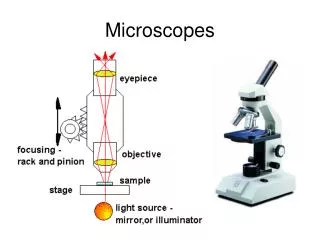
Compound Light microscope • Advantages: can see living specimen • Disadvantage: limited to size and resolution • Uses a 2 lens and a light source • Total magnification is eyepeice x objective • Images
Scanning electron microscope • Advantages of SEM: can see detailed structures and textures of the cell or virus • Disadvantage: can not view live specimen • Creates a vaccum and bombards metal coated specimen with electrons
Transmitting electron microscope • Advantages: see detailed images of interior of cells • Disadvantages: because the specimen is thinly sliced the images do not show active function but a picture in time.
Other images • Potato with and without iodine stain
Other images Blood Human and Amphibian(has a nucleus)
Plant cells Plasmolysis of Elodea, shows all chloroplast and cytoplasm clumped in the center of the cell Cell wall Nucleus. Most of the center looks empty but it is actually a vacuole full of water