Download

1 / 21
210 likes | 450 Views
ABDOMEN AND PELVIS RADIOGRAPHS AND IMAGES: X-RAYS AND ANGIOGRAMS 2007. 7 Arteriogram of Celiac Trunk 1. Common Hepatic artery 2. Gastroduodenal artery 3. Left hepatic artery 4. Right Gastroepiploic artery 5. Splenic artery 6. Left Gastroepiploic artery 7. Gas in loops of bowel.

E N D
ABDOMEN AND PELVIS RADIOGRAPHS AND IMAGES: X-RAYS AND ANGIOGRAMS 2007
7 Arteriogram of Celiac Trunk 1. Common Hepatic artery 2. Gastroduodenal artery 3. Left hepatic artery 4. Right Gastroepiploic artery 5. Splenic artery 6. Left Gastroepiploic artery 7. Gas in loops of bowel
RENAL ARTERIOGRAM 1. External Iliac Artery 2. Common Iliac Artery 3. Aorta 4. Renal Artery 5. Anterior and Posterior Branches of Renal Artery
6 ARTERIOGRAM OF CELIAC TRUNK 1. Celiac Trunk 2. Splenic Artery 3. Common hepatic artery 4. Left Gastric artery 5. Catheter in abdominal aorta 6. Inferior phrenic arteries 7. Spleen
1 Barium swallow upper GI oblique 1. Gas in fundus of stomach 2. Pyloric canal 3. Pylorus 4. First part of duodenum (called duodenal 'bulb' or 'cap') 5. Lesser curvature
11 KIDNEY AND URETER 1. Ureter of Left Kidney 2. Pelvis of Ureter 3. Major Calyx 4. Minor Calyx 5. Lateral border of Right Kidney
KIDNEY AND URINARY BLADDER: CONTRAST IN URINARY SYSTEM 1 Major Calyx 2. Pelvis of ureter 3. Minor Calyx 4. Rib 12 5. Transverse Process of Lumbar vertebra (L4) 6. Sacrum 7. Ureter 8. Urinary bladder 9. bladder 'stone' 10 Lateral border of psoas major
5 CONTRAST IN BILE DUCTS AND GALL BLADDER - see note next slide for technique 1. Tube in 2nd part of duodenum 2. Common bile duct 3. Hepatic ducts 4. Cystic duct 5. Gall bladder 6. Right colic flexure
5 CONTRAST IN BILE DUCTS AND GALL BLADDER Oblique Film; Endoscope inserted into esophagus, through stomach and into 2nd part of duodenum. Contrast material in tube injected into Ampulla of Vater to fill biliary apparatus. (Technique called endoscopic retrograde cholangiopancreaticoagraphy) 1. Tube in 2nd part of duodenum 2. Common bile duct 3. Hepatic ducts 4. Cystic duct 5. Gall bladder 6. Right colic flexure
10 Superior mesenteric Arteriogram gallstones 1. Superior mesenteric artery (SMA) 2. Jejunal branch of SMA 3. Vasa recta (long, characteristic of jejunum) 4. Ileal branch of SMA 5. Arterial arcades - more complex and developed in ileum than in jejunum. Vasa recta are shorter in ileum. 6. Ileocolic artery 7. Right colic artery 8. Marginal artery (of Drummond) 9. Middle colic artery 10. Gall stones
14 Arteriogram of Abdominal Aorta 1. Right renal artery 2. Right common iliac artery 3. Kidney (right) ; note it is lower than left kidney due to liver being on right 4. Abdominal aorta 5. Bifurcation of Aorta (L4)
9 LOWER GI SERIES BARIUM ENEMA 1. Terminal part of ileum 2. Cecum 3. Transverse colon 4. Pelvic (sigmoid) colon 5. Haustra of colon 6. Rectum 7. Descending colon 8. Ascending colon
3 Barium Swallow: Stomach, Duodenum, Jejunum 3 Barium Swallow: Stomach, Duodenum, Jejunum 1. Markings caused by plicae circulares of duodenum (3rd part) 2. Proximal part of jejunum 3. Ascending (4th part) of duodenum 4. Stomach
Intravenous Pyelogram 1. Renal Pelvis 2. Major Calyx 3. Minor Calyx 4. Ureter 5. Ureter crossing pelvic brim (region of potential constriction) 6. Passage of ureter through bladder wall 7. Head of Femur 8. Bladder
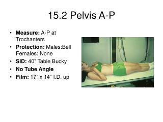
1 X-ray Pelvis and Proximal Femur 1. Anterior Superior Iliac Spine 2. Acetabular rim of pelvis 3. Sacral foramina for Spinal nerve 4. Pubic symphysis 5. Inferior Ramus of Pubis 6. Obturator Foramen 7. Ischium 8. Head of Femur 9. Neck of Femur 10. Greater Trochanter 11. Lesser Trochanter
4 X-ray One Year old Female Thigh Abducted (bones not yet fused) 1. Ilium 2. Pubis 3. Ischium
Uterine Tube Contrast Medium - Hysterosalpingogram (contrast medium injected via vagina to examine patency of uterine tube 1 - Vagina 2. Body of uterus 3. Fundus of uterus 4. Uterine tube 5. Contrast material spillage into peritoneal cavity via patent opening of uterine tube
10 Dye in Penile Urethra Via Catheter 1. Catheter 2. Penile urethra 3. Membranous urethra 4. Urinary bladder 5. Prostatic urethra 5. Radio translucent area = obturator foramen
PELVIS X-RAY AP NORMAL 1. Sacroiliac joint 2. Gas in bowel 4. Superior ramus of pubis 5. Obturator foramen 6. Ischial tuberosity 7. Iliac tubercle 8. Iliac crest 9. Inferior ramus of pubis 10. Pubic symphysis