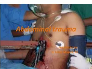
Abdominal trauma
Abdominal trauma. Dr.L.Bahadorzadeh. T he abdomen is frequency injured after both blunt and penetrating trauma. Approximately 25% of all trauma victims will require an abdominal exploration. P hysical examination of the abdomen is unreliable in making intra abdominal injuries.

Abdominal trauma
E N D
Presentation Transcript
Abdominal trauma Dr.L.Bahadorzadeh
The abdomen is frequency injured after both blunt and penetrating trauma. Approximately 25% of all trauma victims will require an abdominal exploration.
Physical examination of the abdomen is unreliable in making intra abdominal injuries. Drugs, alcohol, and head and spinal cord injuries complicate physical examination. It may also be impractical in patients who require general anesthesia for the treatment of other injuries.
Mechanism of injury Blunt trauma secondary to motor vehicle accidents,falls..., remain the most frequent mechanisms of abdominal injury. Penetrating abdominal wounds are usually caused by either gunshot or stab wounds or less shotgun.
Diagnosis ☺The history of the traumatic events ☺ History and physical examination on arrival ☺ Diagnostic modality The test of choice will dependent on the hemodynamic stability of the patient & the severity of associated injuries.
Plain Radiographs ☺ C.X.Ray ☺ Intravenous pyelography ☺ Pelvic Radiography
Diagnostic peritoneal lavage ☺ Indications equivocal pulmonary embolism Unexplained shock or hypotension Altered sensorium(closed head inj,drugs) General anesthesia for extra abdominal procedures Cord injury ☺Contraindications Clear indication for exploratory laparatomy Relative: Previous exploratory laparatomy Pregnancy obesity
…DPL ☺Standard criteria for a positive DPL; Aspiration of at least 10 ml gross blood A bloody lavage effluent A RBC count greater than 100000/mm³ A WBC count greater than 500/mm³ An amylase value greater than 175 IU/dl The detection of bile,bacteria,or food fibers
Sabiston concluded that: Patients sustaining stab wounds can be safely discharge home if the RBC count is less than 1000 provided that they are hemodynamically stable & have no clear indication based on physical examination for operative intervention.
Ultrasound ☺Advantages Non invasive Doesnot reqiure radiation Useful in the resuscitation room or emergency department Can be repeated Used during initial evaluation Low cost ☺Disadvantages Examiner dependent Obesity Gas interposition Lower sensitivity for free fluid <500 ml False negative: retroperitoneal and hollow viscus injuries
Abdominal CT ☺Indications Blunt trauma Hemodynamic stability Normal or unreliable physical examination Mechanism;duodenal and pancreatic trauma ☺Contraindications Clear indication for exploratory laparatomy Hemodynamic instability Agitation Allergy to contrast media
In CT if contrast medium extravasation is seen in minor hepatic and splenic injury an exploratory laparatomy or more recently angiography and embolization are indicated.
The diagnostic approach to penetrating and blunt abdominal trauma differs substantially. As a rule, little preoperative evaluation is required for firearm injuries that penetrate the peritoneal cavity, because the chance of internal injury is over 90% and laparotomy is mandatory
Anterior truncal GSWs between the fourth intercostal space and the pubic symphysis, whose trajectory by x-ray or entrance/exit wound suggests peritoneal penetration, should be operated on.
GSWs to the back or flank are somewhat more difficult to evaluate. If in doubt, it is always safer to explore the abdomen than to equivocate when the depth of penetration is uncertain.
SWs that penetrate the peritoneal cavity are less likely to injure intra-abdominal organs. Anterior and lateral SWs to the trunk should be explored under local anesthesia in the ED to determine whether the peritoneum has been violated. Injuries that do not penetrate the peritoneal cavity do not require further evaluation.
SWs to the flank and back are more difficult to evaluate. Some authorities have recommended a triple-contrast CT to detect occult retroperitoneal injuries.
SWs to the lower chest present a unique diagnostic opportunity. Confirmation of diaphragm penetration by palpation is an indication for laparotomy. when a hole is not palpable, a DPL should be performed. A RBC count in the effluent of more than 10,000 is considered positive when evaluating for a diaphragmatic injury. For RBC counts between 1000 and 10,000, thoracoscopy should be considered.
Blunt abdominal trauma is currently evaluated by US in most major trauma centers, with CT in selected cases to refine the diagnosis. US performed by a surgeon in the ED. US is used in specific anatomic regions (e.g.,Morison's pouch, the left upper quadrant, and the pelvis) to identify free intraperitoneal fluid Although this method is exquisitely sensitive for detecting intraperitoneal fluid collections larger than 250 mL, it is relatively poor for staging solid organ injuries. DPL is still appropriate for patients whose condition cannot be explained by US.
Emergent Abdominal Exploration ☺All abdominal explorations in adults are performed using a long midline incision because of its versatility. Liquid and clotted blood is rapidly evacuate with multiple laparotomy pads and suction. Additional pads are then placed in each quadrant to localize hemorrhage, and the aorta is palpated to estimate blood pressure.
If exsanguinating hemorrhage is encountered upon opening the abdomen, it is usually caused by injury to the liver, aorta, inferior vena cava, or iliac vessels. If the liver is the source, the hepatic pedicle should be immediately clamped (a Pringle maneuver) and the liver compressed posteriorly by tightly packing several laparotomy pads between the hepatic injury and the underside of the right anterior chest wall.(fig.1)
If exsanguinating hemorrhage originates near the midline in the retroperitoneum, direct manual pressure is applied with a laparotomy pad and the aorta is exposed at the diaphragmatic hiatus and clamped. The same approach is used in the pelvis except that the infrarenal aorta can be clamped. venous injuries are not controlled with aortic clamping. A helpful maneuver in these instances is pelvic vascular isolation.(fig2)
For stable patients with large midline hematomas, clamping the aorta proximal to the hematoma is also a wise precaution. Many surgeons take a few moments, once overt hemorrhage has been controlled, to identify obvious sources of enteric contamination and minimize further spillage. This can be accomplished with a running suture or with Babcock clamps.
In blunt trauma, organs that cannot yield to impact by elastic deformation are most likely to be injured. The solid organs,liver, spleen, &kidneys, are representative of this group. For penetrating trauma, organs with the largest surface area are most prone to injury (i.e., the small bowel, liver,and colon).
bullets and knives usually follow straight lines,adjacent structures are commonly injured (e.g., the pancreas and duodenum). Penetrating trauma is not limited by the elastic properties of the tissue, and vascular injuries are far more common.
All abdominal organs are systematically examined by visualization,palpation, or both. Missed injuries: In penetrating trauma failure to explore retroperitoneal structures such as the ascending and descending colons, the second& third portion of the duodenum, and ureters. Injuries of the aorta or vena cava may be temporarily tamponaded by overlying structures. Blunt abdominal injuries of the pancreas, duodenum, bladder, and even the aorta can be overlooked.
Liver Techniques for the temporary control of hemorrhage ☺Manual compression(fig3) ☺ Perihepatic packing (fig3) ☺ The Pringle maneuver (fig3) ☺ Tourniquet ☺ Lin liver clamp
…liver Special techniques for controlling hemorrhage from juxtahepatic venous injuries: ☺ Hepatic vascular isolation with clamps, ☺ The atriocaval shunt (fig4) ☺ The Moore-Pilcherer balloon
If massive venous hemorrhage is seen from behind the liver, and if reasonable hemostasis can be achieved with perihepatic packing, the patient can be transferred to the interventional radiology suite, where hemorrhage from arterial sources are embolized and stents are placed to bridge venous injuries
Numerous methods for the definitive control of hepatic hemorrhage developed. ☺ Minor lacerations may be controlled with manual compression applied directly to the injury site. ☺ electrocautery ☺ Microcrystalline collagen ☺ Topical thrombin ☺ Fibrin glue ☺ Suturing of the hepatic parenchyma (lacerations less than 3 cm in depth)
☺ Venous hemorrhage due to penetrating wounds that traverse the central portion of the liver can be managed by suturing the entrance& exit wounds with horizontal mattress sutures. ☺ Hepatotomy with selective ligation of bleeding vessels is an important technique usually reserved for transhepatic penetrating wounds.(fig5)
☺Hepatic arterial ligation may be appropriate for patients with recalcitrant arterial hemorrhage from deep within the liver.
the subcapsular hematoma; This lesion occurs when the parenchyma of the liver disrupted by blunt trauma, but Glisson's capsule remains intact. The hematoma may be recognized either at the time of the surgery or preoperatively if a CT scan is performed.
Subcapsular hematomas ☺ involving less than 50% of the surface of the liver ☺that are not expanding or ☺ruptured should be left alone orpacked if discovered on exploratory laparotomy.
…the subcapsular hematoma Hematomas that are expanding during an operation may require exploration. These lesions are often caused by uncontrolled arterial hemorrhage, and packing alone may not be successful.
An alternative strategy would be to pack the liver close to the abdomen to control venous hemorrhage and to transport the patient to the angiographic suite for hepatic arteriography and embolization of the bleeding vessel. Ruptured hematomas require exploration and selective ligation, with or without packing.
☺ Resectional debridement is indicated for the removal of peripheral portions of nonviable hepatic parenchyma. The mass of tissue removed should rarely exceed 25% of the liver. ☺ anatomic lobectomy
Drain are not necessarily for minor laceration. They should be used if bile is seen oozing from the liver and in most patient with deep central injuries.
The complications following significant hepatic trauma ; ☺Hemorrhage ☺Infections ☺Bilomas ☺ Biliary fistulas ☺arterialpseudoaneurysms ☺Biliovenous fistulas
Non operative treatment The classic criteria ☺ hemodynamic stability ☺Normal mental status ☺Absence of a clear indication for laparatomy;peritoneal sign ☺Low grade liver injury ☺ Transfusion requirment of less than 2 units