Download
1 / 15
951 likes | 6.22k Views
Ultrasound abdomen atlas. A&E medical meeting 28/07/2011 Dr. David Tran. FAST : F ocused A ssessment for S onographic evaluation of T rauma patients. four transducer positions (1) pericardial area (2) right upper quadrant (3) left upper quadrant (4) pelvis.

E N D
Ultrasound abdomen atlas A&E medical meeting 28/07/2011 Dr. David Tran
FAST: Focused Assessment for Sonographic evaluation of Trauma patients four transducer positions • (1) pericardial area • (2) right upper quadrant • (3) left upper quadrant • (4) pelvis.
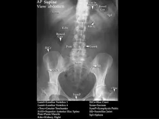
Just below the diaphragm, the vena cava is surrounded by liver tissue.The aorta lies directly behind the gastroesophagealjunction, often making the vessel more difficult to scan • 20: Right liver • 21: Left liver • 10: veina cava • 1 Aorta • 70:Stomack • 90: Thoracic vertebra
10: Veina Cava • 14: Right Renal vein • 20: Right liver • 21: Left liver • 30: Gallbladder • 60: Right kidney • 90: Spinal column • 95: Psoas muscle
20: Liver 32: Gallbladder 10: Veina Cava 14: Right renal vein 76: Duodenum 92: Acoustic shadow
Left upper abdomen Scan(longitudinal view) • 50: Spleen • 43: tail of pancreas • 61: left kidney • 15: Left renal vein and splenic vein (18)
Left longitudinal flank scan 50: Slpeen 61: Left kidney 69: Supra-renal gland 94: Acoustic artifacts
Lower abdominal longitudinal scan • 80: Bladder • 85: Uterus • 89: Rectum
Hypogastric region, longitudinal scan(women) 80: Bladder 86: Uterus 89: rectum (94: Artefact)