Able to display extremely small changes in the brain
80 likes | 176 Views
Learn about advanced neuroimaging techniques like fMRI, PET, and SPECT scans to visualize brain activity during tasks. These non-invasive methods offer detailed insights into brain function and structure, aiding in diagnosing brain disorders. Discover their benefits and limitations.

Able to display extremely small changes in the brain
E N D
Presentation Transcript
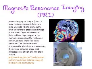
A neuroimaging technique (like a CT scan) that uses magnetic fields and radio waves to vibrate atoms in the brain’s neurons to produce and image of the brain. These vibrations are detected by a huge magnet in the chamber surrounding the motionless person, and are channeled into a computer. The computer then processes the vibrations and assembles them into a coloured image that indicates areas of high and low brain activity. More sensitive than a CT and provides a clearer and more detailed image of the brain and its structures.
Able to display extremely small changes in the brain • Can more clearly distinguish the different between brain tissue that is cancerous and non cancerous. • Can be used to diagnose a wide range of brain disorders • Non invasive and harmless • It does not use X-rays or radioactive substances • Does not provide information about the activity of the brain, only shows the anatomy of the brain. • Costly • Can not be used on people who have internal metallic devices
A neuroimaging technique that is able to provide a 3D image of the brains activity and function while the participant is completing various tasks. PET scans track blood flow around the brain while it is working during usually simple task such as talking or counting and as it is assumed that the brain areas that require increased blood flow have increased neural activity, radiologists are able to see what parts of the brain are working during specific tasks. Prior to the PET scan, a harmless radioactive substance is injected into the participants blood vessels, this substance travels to the brain and can be followed as it emits radioactive signals. These signals are detected and recorded onto the PET computer. A PET scan looks like a coloured ‘map’ of the brains activity with different colours indicating the areas of greatest and least activity,
Highly effective in diagnosing abnormalities in the brain • An area of brain deterioration can be easily identified (as shown the the picture on the previous slide) • Is able to provide information on both function and structure • Able to identify brain areas that are active and inactive in normal people during everyday activities • Non invasive and harmless • Researchers cannot determine whether an active brain area is actually involved in the mental process or behavior under investigation • Radioactivity decays rapidly therefore PET scans are limited to study relatively short tasks • Doesn’t pick up on rapid progression • Costly
A variation of the PET scan technique Uses a longer lasting radioactive tracer and a scanner to record data that a computer uses to construct two or three dimensional images of active brain regions. The similarity of image processing between a SPECT and a CT allow the images to be combined when a better resolution is required. The tracer decays a lot slower so participants are able to be given longer lasting tasks in research studies.
Cheaper then a PET scan, so when suitable to do so, researchers tend to use a SPECT over a PET scan • Non invasive • Radioactive dye lasts longer so tasks can be longer • Show function and structure • Image is not as accurate and clear as a PET scan image as it has a lower resolution • Unable to observe rapid changes in brain activity for functional research
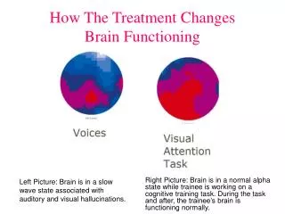
A neuroimaging technique that enables the identification of brain areas that are particularly active during a given task by detecting changes in oxygen levels in the blood flowing through the brain. A computer analyses the blood oxygen levels in the area, and creates an image with colour variations, like a PET and SPECT with the colours reflecting the level of activity in that part of the brain. An fMRI is able to take numerous pictures of the brain in rapid succession and can therefore detect brain changes as they occur from moment to moment and fMRI images of brain structures and activity are more highly detailed and precise.
Does no require exposure to radiation • Enables detailed images of the functioning brain while a person performs a task requiring mental processes or behavioral responses. • More detailed then SPECT and PET scan images • Easy to interpret with the use of colour coding • There is no scope for suggesting cause-effect relationships between active and inactive areas and mantel processes and behaviors under investigation • Costly