Download
1 / 48
510 likes | 708 Views
Structural Changes in the Brain of Addicts. Emery Neuroscience Center 5340 N Federal Hwy, Ste. 205 Lighthouse Point, FL 33064 Tel: 954-771-8300 . Waden E. Emery III M.D. FAAN Asst. Clinical Professor Neurology University of Miami. Recovery and Community: It Takes a Village

E N D
Structural Changes in the Brain of Addicts Emery Neuroscience Center 5340 N Federal Hwy, Ste. 205 Lighthouse Point, FL 33064 Tel: 954-771-8300 Waden E. Emery III M.D. FAAN Asst. Clinical Professor Neurology University of Miami Recovery and Community: It Takes a Village September 27th 2013
Overview • Current Imaging Techniques (primarily research) • Anatomical Regions believed to be involved in Addiction • Anomalies seen in the addicted brains • Neurotransmitters involved in addiction • Genetic variations alter anomalies of brain anatomy • Treatment strategies
Overview • Current Imaging Techniques (primarily research) • Anatomical Regions believed to be involved in Addiction • Anomalies seen in the addicted brains • Neurotransmitters involved in addiction • Genetic variations alter anomalies of brain anatomy • Treatment strategies
Brain Imaging Techniques • Structural Magnetic Resonance Imaging (MRI) • Functional Magnetic Resonance Imaging (fMRI) • Magnetic Resonance Spectroscopy (MRS) • Positron Emission Tomography (PET) • Single Photon Emission Computed Tomography (SPECT)
Structural MRI (MRI) • Map Tissue morphology and composition
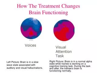
Functional MRI (fMRI) • Visualize changes in oxygenation and blood flow associated with brain activities
Magnetic resonance spectroscopy (MRS) • Measure cerebral metabolism, physiological processes involving specific brain chemicals; detect drug metabolites
Positron emission tomography (PET) • Quantify biochemical and pharmacological processes • Glucose metabolism • Drug distribution and kinetics • Receptor ligand interaction • Enzyme Targeting
Single photon emission computed tomography (SPECT) • Measure receptor ligand interaction • Measure physiological function • Measure biochemical processes • Measure pharmacological processes
Overview • Current Imaging Techniques (primarily research) • Anatomical Regions believed to be involved in Addiction • Anomalies seen in the addicted brains • Neurotransmitters involved in addiction • Genetic variations alter anomalies of brain anatomy • Treatment strategies
Anatomical Regions Involved in Addiction • Orbitofrontal Cortex (OFC) • Ventral Tegmental Area (VTA) • Prefrontal Cortex • Frontal Cortex • Parietal Cortex • Basal Ganglia • Cingulate Gyrus • Nucleus Accumbens (NAc) • Amygdala • Hippocampus
Functional Correlates • The Ventral Tegmental Area (VTA) & Nucleus Accumbens (NAc) are key components of the brain’s reward system • The VTA, NAc, amygdala, and hippocampus are major components of the limbic system, which coordinates drives, emotions, and memories
Overview • Current Imaging Techniques (primarily research) • Anatomical Regions believed to be involved in Addiction • Anomalies seen in the addicted brains • Neurotransmitters involved in addiction • Genetic variations alter anomalies of brain anatomy • Treatment strategies
Structural MRI Changes • Addictive drugs cause volume and tissue composition changes in the Frontal cortex • These changes are likely associated with abusers’ cognitive and decision making problems. • Frontal Lobes and Prefrontal Lobes are decreased size in drug users
Structural MRI Changes • Cocaine dependent individuals have enlargement of the basal ganglia compared to normals • Methamphetamine abusers had severe gray matter deficits in the cingulate, limbic, and paralimbic cortices. • Methamphetamine abusers had smaller hippocampi than normals (hippocampi are a key site for memory storage and volume decrements correlated with poorer performance on a word recall test
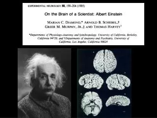
Structural MRI Changes • The amygdala, a brain structure that helps shape our emotional responses to experiences is relatively small in children of alcoholics
Structural MRI Changes • Methamphetamine Reduces Grey Matter
Functional MRI (fMRI) Changes • fMRI studies reveal changes resulting from the ratio of oxygenated to deoxygenated hemoglobin (During a task, Oxygen consumption is raised in specific areas of the brain and therefore the ratio of oxygenated to deoxygenated hemoglobin changes) • When given cocaine in a fMRI machine, the drug rush correlates with higher activity (Oxygen consumption) in the caudate, cingulate, and lateral prefrontal cortex
Functional MRI (fMRI) Changes • Reports of craving commenced when the euphoria subsided and persisted as long as a different set of brain areas including the NAc remained activated • Other studies suggested higher activation in the NAc, inferior frontal/orbitofrontal gyrus and anterior cingulate • Activation of the anterior cingulate cortex, an area associated with emotional processing persisted while cocaine-addicted subjects watched videotapes containing cocaine-accociated cues
Functional MRI(fMRI) Changes • The Brains Response to Cocaine Cues-Anterior Cingulate activated in Cocaine users
Magnetic Resonance Spectroscopy (MRS) Changes • MRS scans reveal the location and concentrations of target chemicals in brain tissue • Two that have been studied are N-Acetylaspartate (NAA) which has been used as a gauge of neuronal cell health and myoinositol, which is present in support cells, glia • Choline compounds which are involved in turnover of cell membranes and creatinine compounds which are important for cells’ energy maintenance
Magnetic Resonance Spectroscopy (MRS) Changes • Methamphetamine users have reduced NAA concentrations in basal ganglia and frontal white matter—measures of NAA have correlated with measures of cognitive function • Cocaine users have decreased NAA levels, suggesting neuron damage, as well as elevated creatine and myoinositol levels reflecting increased glial cell activity or inflammation
PET & SPECT Changes • PET & SPECT scans display the distribution of a labeled compound, called a radiotracer • PET & SPECT vary with respect to the type of tracer • Dopamine is highly concentrated in the striatum, which forms part of the brain’s reward system—its levels determine how much pleasure we derive from our experiences and helps us focus our attention on what is important
PET & SPECT Changes • PET & SPECT studies have established that cocaine, amphetamine, and methylphenidate, when given intravenously produce their highs by massively increasing the volume of dopamine in the striatum • Further studies have shown that any drug’s abuse liability depends both on the size of the dopamine spike it produces and the rapidity with which dopamine rises and falls back to normal levels
PET & SPECT Changes • The “high” experienced correlates with uptake of cocaine in the striatum
Cocaine leads to a build up of dopamine in the synaptic cleft
PET & SPECT Changes • Chronic exposure reduces the availability of dopamine transporters, suggesting a loss of dopamine cells • Cocaine and methamphetamine reduce cellular activity in the orbitofrontal cortex (OFC) a brain area used to make strategic rather than impulsive decisions • Traumatic brain injuries to the OFC lead to aggressiveness, poor judgment of future consequences and the inability to inhibit inappropriate responses
PET & SPECT Changes • Cocaine abusers have decreased metabolism in the OFC (Right) compared to normals (Left)
PET & SPECT Changes • striatum of the healthy control (left) is largely red, indicating the highest level of receptor availability, while that of the cocaine abuser has little red.
PET & SPECT Changes • Cocaine users decreased judgment has correlated with decreased metabolism in the OFC • Abusers of alcohol, cocaine, heroin and methamphetamine all have reduced levels of brain dopamine receptors
PET & SPECT Changes • Smokers have decreased MAO, enzyme that metabolizes dopamine
PET & SPECT Changes • Smokers have low levels of MAO (monoamine oxidase) which breaks down dopamine • Releases hydrogen peroxide, a potential source of free radicals that can damage nerve cells • MAO-inhibiting chemical compounds have been isolated from tobacco and shown to have a protective action in rodent model of Parkinson’s disease
Overview • Current Imaging Techniques (primarily research) • Anatomical Regions believed to be involved in Addiction • Anomalies seen in the addicted brains • Neurotransmitters involved in addiction • Genetic variations alter anomalies of brain anatomy • Treatment strategies
Genetic Variations • Genetic variations have been documented in individuals with respect to their response to amphetamine • Genetic variation could increase sensitivity to stress and heighten vulnerability to drug abuse • Current hypothesis is that individuals with low levels of dopamine receptors, either genetically or as a result of experiences, have a higher risk of abuse and addiction.
Genetic Variations • Individuals with fewer dopamine receptors obtain less than normal amounts of dopamine-mediated pleasure from ordinary activities and accomplishments and therefore are highly susceptible to wanting to repeat the euphoria that occurs when drugs massively increase dopamine in the brain
Overview • Current Imaging Techniques (primarily research) • Anatomical Regions believed to be involved in Addiction • Anomalies seen in the addicted brains • Neurotransmitters involved in addiction • Genetic variations alter anomalies of brain anatomy • Treatment strategies
Treatment Strategies • Medications are being developed that will produce only a modest dopamine spike causing susceptible individuals to experience pleasure normally • MAO-B inhibitors and other medications fitting this criterion have been used to treat smoking addiction
Treatment Strategies • Compounds that enhance the neurotransmitter gamma-aminobutyric acid (GABA) which has been shown to inhibit dopamine-releasing cells’ response to drug related cues • Other drugs are currently under study that modify the dopamine-receiving cells and thereby attenuate the reinforcing effects of abused drugs
Treatment Strategies • Drugs are currently under study that cause only a modest dopamine spike instead of a sharp spike---treatment of heroin addiction with methadone and buprenorphine exemplify this approach • Functional MRI studies of men entering treatment for methamphetamine addiction while they made decisions during a psychological test showed two patterns and predicted with 90 percent accuracy which of the men would relapse within 1 to 3 years after completing treatment---those who relapsed had less activity in the prefrontal lobe
Summary • Imaging techniques have defined the regions of the brain that are altered when exposed to various chemicals (drugs) in addicted individuals • Areas of the brain are those involved with reward, coordination of drives, emotions, and memories • The “high” from drug usage correlates with the neurotransmitter spike dopamine in specific areas of the brain
Summary • Medications are currently used and being developed to alter the dopamine spike • Genetic variations to the dopamine spike have been identified • Imaging techniques may help to predict potential recidivism rates
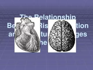
Lessons from Neurology • Dopamine agonists in Parkinson’s Disease can cause obsessive/compulsive behavior, eg, gambling, sexual addictions---now carry a warning from FDA • Cocaine users have a significantly increased risk of stroke, cerebral vasculitis • Alcoholics can develop Wernicke and or Korsakoff’s Syndromes due to vitamin deficiency
Lessons from Neurology • Delerium Tremens from alcohol/drug withdrawal leads to seizures----my number one consult while taking call at Broward Health Imperial Point • Parkinson’s Disease from ingestion of MPTP, a synthetic form of heroin • Polyneuropathy and focal neurapraxia from drug ingestion
Lessons from Neurology • Cerebellar Degeneration from long term alcohol abuse • Alcohol Dementia • Fetal Alcohol Syndrome • Myopathy-Alcohol • Tremor associated with Amphetamines • Decrease in Memory/Higher Cortical Functions/Seizures associated with benzodiazepines