Download
1 / 32
330 likes | 516 Views
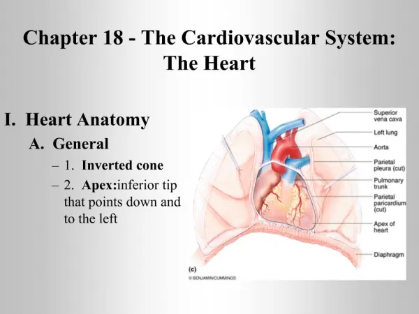
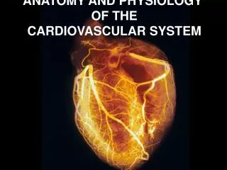
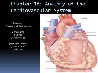
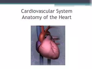
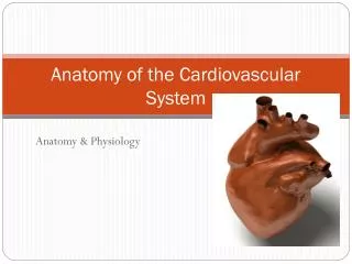
Chapter 18 Anatomy of the Cardiovascular System. Location of the heart . Lies in the mediastinum, behind the body of the sternum between the points of attachment of ribs 2 through 6; approximately two thirds of its mass is to the left of the midline of the body and one third to the right

E N D
Location of the heart • Lies in the mediastinum, behind the body of the sternum between the points of attachment of ribs 2 through 6; approximately two thirds of its mass is to the left of the midline of the body and one third to the right • Posteriorly the heart rests on the bodies of thoracic vertebrae 5 through 8 • Apex lies on the diaphragm, pointing to the left • Base lies just below the second rib • Boundaries of the heart are clinically important as an aid in diagnosing heart disorders
Coverings of the heart • Structure of the heart coverings • Pericardium • Fibrous pericardium—tough, loose-fitting inextensible sac • Serous pericardium—parietal layer lies inside fibrous pericardium, and visceral layer (epicardium) adheres to outside of the heart; pericardial space with pericardial fluid separates the two layers • Function of the heart coverings—provides protection against friction
Structure of the heart • Wall of the heart—made up of three distinct layers: • Epicardium—outer layer of heart wall • Myocardium—thick, contractile middle layer of heart wall; compresses the heart cavities, and the blood within them,with great force • Endocardium—delicate inner layer of endothelial tissue
Structure of the heart (cont.) • Chambers of the heart—divided into four cavities with the right and left chambers separated by the septum: • Atria • Two superior chambers, known as “receiving chambers,” because they receive blood from veins • Atria alternately contract and relax to receive blood and then push it into ventricles • Myocardial wall of each atrium is not very thick, because little pressure is needed to move blood such a small distance • Auricle—earlike flap protruding from each atrium • Ventricles • Two lower chambers, known as “pumping chambers,” because they push blood into the large network of vessels • Ventricular myocardium is thicker than myocardium of the atria, because great force must be generated to pump blood a large distance; myocardium of left ventricle is thicker than the right, because it must push blood much further
Structure of the heart (cont.) • Valves of the heart—mechanical devices that permit the flow of blood in one direction only • Atrioventricular (AV) valves—prevent blood from flowing back into the atria from the ventricles when the ventricles contract • Tricuspid valve (right AV valve)—guards the right atrioventricular orifice; free edges of three flaps of endocardium are attached to papillary muscles by chordae tendineae • Bicuspid, or mitral, valve (left AV valve)—similar in structure to tricuspid valve except only two flaps present
Structure of the heart (cont.) • Valves of the heart (cont.) • Semilunar (SL) valves—half moon–shaped flaps growing out from the lining of the pulmonary artery and aorta; prevent blood from flowing back into ventricles from aorta and pulmonary artery • Pulmonary semilunar valve—at entrance of pulmonary artery • Aortic semilunar valve—at entrance of aorta
Structure of the heart (cont.) • Blood supply of heart tissue • Coronary arteries—myocardial cells receive blood from right and left coronary arteries • First branches to come off aorta • Ventricles receive blood from branches of both right and left coronary arteries • Each ventricle receives blood only from a small branch of corresponding coronary artery • Most abundant blood supply goes to myocardium of left ventricle • The right coronary artery is dominant in approximately 50% of all hearts and the left in about 20%; in approximately 30%, neither coronary artery is dominant • Few anastomoses exist between the larger branches of the coronary arteries
Structure of the heart (cont.) • Blood supply of heart tissue (cont.) • Veins of the coronary circulation • As a rule, veins follow a course that closely parallels that of coronary arteries • After going through cardiac veins, blood enters coronary sinus to drain into right atrium • Several veins drain directly into right atrium
Structure of the heart (cont.) • Conduction system of the heart—comprising the sinoatrial (SA) node, atrioventricular (AV) node, AV bundle, and Purkinje fibers; made up of modified cardiac muscle (Figure 18-11) • Sinoatrial node (SA node or pacemaker)— hundreds of cells in right atrial wall near opening of superior vena cava • Atrioventricular node (AV node)—small mass of special cardiac muscle in right atrium along lower part of interatrial septum • Atrioventricular bundle (AV bundle or bundle of His) and Purkinje fibers • AV bundle originates in AV node, extends by two branches down the two sides of the interventricular septum, and continues as Purkinje fibers • Purkinje fibers extend out to papillary muscles and lateral walls of ventricles
Structure of the heart (cont.) • Nerve supply of the heart • Cardiac plexuses—located near arch of aorta, made up of the combination of sympathetic and parasympathetic fibers • Fibers from cardiac plexus accompany right and left coronary arteries to enter the heart • Most fibers end in the SA node, but some end in the AV node and in the atrial myocardium • Sympathetic nerves—accelerator nerves • Vagus fibers—inhibitory, or depressor, nerves
Blood Vessels • Types of blood vessels • Arteries • Carry blood away from heart—all arteries except pulmonary artery carry oxygenated blood • Elastic arteries—largest in body • Examples: aorta and its major branches • Able to stretch without injury • Accommodate surge of blood when heart contracts and able to recoil when ventricles relax
Blood Vessels • Arteries (cont.) • Muscular (distributing) arteries • Smaller in diameter than elastic arteries • Muscular layer is thick • Examples: brachial, gastric, superior mesenteric • Arterioles (resistance vessels) • Smallest arteries • Important in regulating blood flow to end-organs • Metarterioles • Short connecting vessel between true arteriole and 20 to 100 capillaries • Encircled by precapillary sphincters • Distal end called thoroughfare channel, which is free of precapillary sphincters
Blood Vessels • Types of blood vessels (cont.) • Capillaries—primary exchange vessels • Microscopic vessels • Carry blood from arterioles to venules—together, arterioles, capillaries and venules constitute the microcirculation • Not evenly distributed—highest numbers in tissues with high metabolic rate; may be absent in some “avascular” tissues such as cartilage
Blood Vessels • Types of capillaries • True capillaries—receive blood flowing from metarteriole with input regulated by precapillary sphincters • Continuous capillaries • Continuous lining of endothelial cells • Openings called intercellular clefts exist between adjacent endothelial cells • Fenestrated capillaries • Have both intercellular clefts and “holes” or fenestrations through plasma membrane to facilitate exchange functions • Sinusoids • Large lumen and tortuous course • Absent or incomplete basement membrane • Very porous—permit migration of cells into or out of vessel lumen
Blood Vessels • Types of blood vessels (cont.) • Veins • Carry blood toward the heart • Act as collectors and as reservoir vessels; called capacitance vessels
Blood Vessels • Structure of blood vessels • Layers • Tunica adventitia—found in arteries and veins • Tunica media—found in arteries and veins • Tunica intima—found in all blood vessels; only layer present in capillaries
Blood Vessels • Structure of blood vessels (cont.) • “Building blocks” commonly present • Lining endothelial cells • Only lining found in capillary • Line entire vascular tree • Provide a smooth luminal surface—protects against intravascular coagulation • Intercellular clefts, cytoplasmic pores, and fenestrations allow exchange to occur between blood and tissue fluid • Capable of secreting a number of substances • Capable of reproduction
Blood Vessels • “Building blocks” commonly present (cont.) • Collagen fibers • Exhibit woven appearance • Formed from protein molecules that aggregate into fibers • Visible with light microscope • Have only a limited ability to stretch (2% to 3%) under physiological conditions • Function to strengthen and keep lumen of vessel open
Blood Vessels • “Building blocks” commonly present (cont.) • Elastic fibers • Composed of insoluble protein called elastin • Form highly elastic networks • Fibers can stretch over 100% under physiological conditions • Play important role in creating passive tension to help regulate blood pressure throughout cardiac cycle • Smooth muscle fibers • Present in all segments of vascular system except capillaries • Most numerous in elastic and muscular arteries • Exert active tension in vessels when contracting
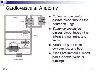
Major Blood Vessels • Circulatory routes • Systemic circulation—blood flows from the left ventricle of the heart through blood vessels to all parts of the body (except gas exchange tissues of lungs) and back to right atrium • Pulmonary circulation—venous blood moves from right atrium to right ventricle to pulmonary artery to lung arterioles and capillaries where gases are exchanged; oxygenated blood returns to left atrium via pulmonary veins; from left atrium, blood enters left ventricle
Major Blood Vessels • Systemic circulation • Systemic arteries • Main arteries give off branches, which continue to rebranch, forming arterioles and then capillaries • End-arteries—arteries that eventually diverge into capillaries • Arterial anastomosis—arteries that open into other branches of the same or other arteries; incidence of arterial anastomoses increases as distance from the heart increases • Arteriovenous anastomoses or shunts occur when blood flows from an artery directly into a vein
Major Blood Vessels • Systemic circulation (cont.) • Systemic veins • Veins are the ultimate extensions of capillaries; unite into vessels of increasing size to form venules and then veins • Large veins of the cranial cavity are called dural sinuses • Veins anastomose as do arteries • Venous blood from the head, neck, upper extremities, and thoracic cavity (except lungs) drains into superior vena cava • Venous blood from thoracic organs drains directly into superior vena cava or azygos vein • Hepatic portal circulation • Veins from the spleen, stomach, pancreas, gallbladder, and intestines send their blood to the liver via the hepatic portal vein • In the liver, venous blood mingles with arterial blood in the capillaries and is eventually drained from liver by hepatic veins that join the inferior vena cava • Venous blood from lower extremities and abdomen drains into inferior vena cava
Major Blood Vessels • Fetal circulation • Basic plan of fetal circulation—additional vessels needed to allow fetal blood to secure oxygen and nutrients from maternal blood at the placenta (Figure 18-31) • Two umbilical arteries—extensions of internal iliac arteries; carry fetal blood to placenta • Placenta—attached to uterine wall, where exchange of oxygen and other substances between the separated maternal and fetal blood occurs (Figure 18-30) • Umbilical vein—returns oxygenated blood from placenta to fetus; enters body through umbilicus and goes to undersurface of liver, where it gives off two or three branches and then continues as ductus venosus • Ductus venosus—continuation of umbilical vein, drains into inferior vena cava • Foramen ovale—opening in septum between right and left atria • Ductus arteriosus—small vessel connecting pulmonary artery with descending thoracic aorta
Major Blood Vessels • Fetal circulation (cont.) • Changes in circulation at birth (compare Figures 18-31 and 18-32) • When umbilical cord is cut, the two umbilical arteries, the placenta and the umbilical vein no longer function • Umbilical vein within the baby’s body becomes the round ligament of the liver • Ductus venosus becomes the ligamentum venosum of the liver • Foramen ovale—functionally closed shortly after a newborn’s first breath and pulmonary circulation is established; structural closure takes approximately 9 months • Ductus arteriosus—contracts with establishment of respiration, becomes ligamentum arteriosum
Cycle of Life: Cardiovascular Anatomy • Birth—change from placenta-dependent system • Heart and blood vessels maintain basic structure and function from childhood through adulthood • Exercise thickens myocardium and increases supply of blood vessels in skeletal muscle tissue • Adulthood through later adulthood—degenerative changes • Atherosclerosis—blockage or weakening of critical arteries • Heart valves and myocardial tissue degenerate—reduces pumping efficiency