Download

1 / 21
210 likes | 238 Views
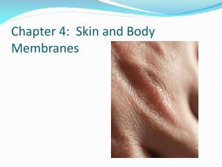
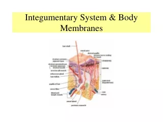
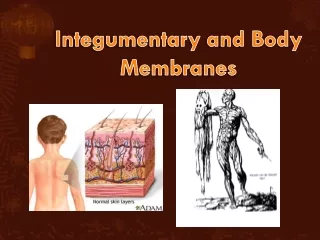
Chapter 4 The Integumentary System and Body Membranes. HAP Susan Chabot Lemon Bay High School. Classification of Body Membranes. Epithelial Membranes Cutaneous Membranes = The Skin Mucous Membranes Serous Membranes Connective Tissue Membranes Synovial Membranes. Cutaneous Membranes.

E N D
Chapter 4The Integumentary System and Body Membranes HAP Susan Chabot Lemon Bay High School
Classification of Body Membranes • Epithelial Membranes • Cutaneous Membranes = The Skin • Mucous Membranes • Serous Membranes • Connective Tissue Membranes • Synovial Membranes
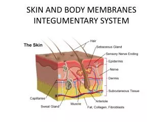
Cutaneous Membranes • The Skin
Mucous Membranes • Lines body cavities that open to the outside, such as THOSE OF HOLLOW ORGANS of the RESPIRATORY, DIGESTIVE, URINARY, REPRODUCTIVE systems. • Adapted for ABSORPTION or SECRETION • The lining of the RESPIRATORY and DIGESTIVE tracts secrete large amounts ofProtective, lubricating mucus.
Serous Membranes • Lines body cavities that are closed to the exterior. • Occur in pairs • Parietal: lines the wall of the ventral body cavity. • Visceral: covers the outside of the organ in that cavity. • Serous Fluid: thin, watery fluid secreted by the membranes. • Function/Importance of Serous Fluid: allows the organs to slide easily across one another without creating friction.
Synovial Membranes • Composed of soft connective tissue • Contain NO epithelial cells • Line the capsules surrounding joints • Provides smooth, lubricating fluid to cushion during muscle movement.
Cutaneous Membranes: The Skin Functions • Protection from • Mechanical Damage • Chemical Damage • Bacterial Damage • Ultraviolet Radiation • Temperature Damage • Desiccation/Drying out • Temperature control: sweat and goosebumps • Excretion of urea and uric acid • Synthesis of vitamin D
Appendages of Skin/Glands Accessory Structures that provide specific functions to the Integumentary System. • Cutaneous Glands – Exocrine • Sebaceous Glands/OIL GLANDS • Sebum – antimicrobial properties and keeps skin soft.
Appendages of Skin/Glands • Sudoriferous Glands/SWEAT GLANDS • Eccrine/Merocrine – produces watery sweat for evaporative cooling to control body temperature. • Widely distributed all over the body • Sweat – mostly salt water and other waster materials • Apocrine – produces oily sweat • Located in Axilla and Groin • Becomes active during puberty
EPIDERMIS Composed of 5 Layers/strata • Innermost – Stratum basale = dividing layer • S. spinosum • S. granulosum • S. lucidum • Outermost – S. corneum CHARACTERISTICS • Avascular – lacks rich blood supply • Most cells are keratinocytes that produce KERATIN • Average person sheds 40 pounds of skin in a lifetime • Completely new epidermis every 25 – 45 days • Melanin production is performed by MELANOCYTES in the s. basale during UV exposure.
DERMIS • AKA hide • Strong and elastic • Made of dense fibrous connective tissue
2 Regions of the Dermis Papillary layer = Most superficial layer of the dermis • Dermal papillae attaches DERMIS to s. basale • Forms fingerprints which provide gripping action for fingers • Provides nourishment to the s. basale to keep those cells dividing. Reticular layer = Deepest layer of the dermis • Contains blood vessels, sweat and oil glands, and pressure receptors. • Phagocytes prevent bacteria from entering interior of body • NONSPECIFIC • Collagen (protein) provides toughness for attachment • Elastin (protein) provides flexibility and elasticity. • Maintains body temperature by promoting sweat production and release and contraction of muscles for goosebumps.
HYPODERMIS • Made of adipose tissue = FAT • Deepest layer of the skin • Anchors skin to underlying tissues/organs. • Provides shock absorption and insulation
NORMAL Skin Pigments 3 main pigments contribute to skin color • Melanin: yellow, reddish brown, dark brown, black. Protection from UV light. • Carotene: orange-yellow fat soluble vitamin that can be stored in adipose tissue. • Hemoglobin: red; found in red blood cells. The more blood in the area, the more red the skin appears.
ABNORMAL Skin Coloration Cyanosis: blue cast to skin due to low blood oxygen levels or poor circulation. Erythema: redness due to blushing, inflammation, fever, high blood pressure Pallor: pale skin due to anemia (low RBC), low blood pressure, loss of blood flow. Jaundice: yellow cast due to bile build up; signifies liver damage Bruises: loss of blood due to injury. Color changes to bruise occur as pigments are broken down.
Albinism Erythema Jaundice Cyanosis Jaundice Vitligo