Download
1 / 61
630 likes | 1.43k Views
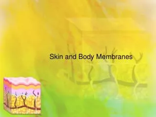
Chapter 4. Skin and body Membranes. Body Membranes. Function of body membranes Cover body surfaces Line body cavities Form protective sheets around organs. Classification of Body Membranes. Epithelial membranes Cutaneous membranes Mucous membranes Serous membranes

E N D
Chapter 4 Skin and body Membranes
Body Membranes • Function of body membranes • Cover body surfaces • Line body cavities • Form protective sheets around organs
Classification of Body Membranes • Epithelial membranes • Cutaneous membranes • Mucous membranes • Serous membranes • Connective tissue membranes • Synovial membranes
Cutaneous Membrane • Cutaneous membrane = skin • Dry membrane • Outermost protective boundary • Superficial epidermis is composed of keratinized stratified squamous epithelium • Underlying dermis is mostly dense connective tissue
Mucous Membranes • Surface epithelium type depends on site • Stratified squamous epithelium (mouth, esophagus) • Simple columnar epithelium (rest of digestive tract) • Underlying loose connective tissue (lamina propria) • Lines all body cavities that open to the exterior body surface
Serous Membranes • Surface is a layer of simple squamous epithelium • Underlying layer is a thin layer of areolar connective tissue • Lines open body cavities that are closed to the exterior of the body • Serous membranes occur in pairs separated by serous fluid • Visceral layer covers the outside of the organ • Parietal layer lines a portion of the wall of ventral body cavity
Serous Membranes Figure 4.1d
Serous Membranes • 3 primary serous membranes • Peritoneum • Abdominal cavity • Pleura • Around the lungs • Pericardium • Around the heart
Serous Membranes Figure 4.1
Connective Tissue Membrane • Synovial membrane • Connective tissue only • Lines fibrous capsules surrounding joints • Secretes a lubricating fluid
The membrane that covers the surface of the heart is the _________________. • parietal peritoneum • visceral pleura • visceral pericardium • parietal pericardium • synovial membrane
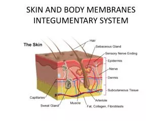
Integumentary System • Skin (cutaneous membrane) • Skin derivatives – Accessory organs • Sweat glands • Oil glands • Hair • Nails • Why is the skin classified as an organ?
Skin Functions Table 4.1 (1 of 2)
Skin Functions Table 4.1 (2 of 2)
Skin Structure • Epidermis—superficial layer • Stratified squamous epithelium • Often keratinized (hardened by keratin) • Keratinization - process by which cytoplasm fills with keratin, a water-proofing protein • Keratinocytes
Skin Structure • Epidermis—superficial layer • Avascular • Deepest cell layers receive nutrients from dermal blood vessels • As cells push toward the surface, they move away from nutrient supply and die • Dermis—deep layer • Dense connective tissue, blood vessels, nerve endings, glands
Skin Structure Figure 4.3
Skin Structure • Subcutaneous tissue (hypodermis) is deep to dermis • Not part of the skin • Anchors skin to underlying organs • Composed mostly of adipose tissue
Layers of the Epidermis • Stratum basale (stratum germinativum) • Deepest layer of epidermis • Lies next to dermis • Cells undergoing mitosis • Daughter cells are pushed upward to become the more superficial layers • Stratum spinosum • Stratum granulosum
Layers of the Epidermis • Stratum lucidum • Formed from dead cells of the deeper strata • Occurs only in thick, hairless skin of the palms of hands and soles of feet • Stratum corneum • Outermost layer of epidermis • Shingle-like dead cells are filled with keratin (protective protein prevents water loss from skin)
Layers of the Epidermis • Summary of layers from most superficial to deepest • Stratum corneum • Stratum lucidum (thick, hairless skin only) • Stratum granulosum • Stratum spinosum • Stratum basale
Melanin • Pigment (melanin) produced by melanocytes • Melanin absorbs UV radiation preventing DNA mutations of skin cells • Color is yellow to brown to black (genetics) • Melanocytes are mostly in the stratum basale • Everyone has the same number • Produce different amounts and sizes of pigment granule (genetics) • Transfers melanin to keratinocytes • Sunlight stimulates melanin production
Dermis • Two layers • Papillary layer (superficial dermal region) • Projections called dermal papillae • Some contain capillary loops • Other house pain receptors and touch receptors • Reticular layer (deepest skin layer) • Blood vessels • Sweat and oil glands • Deep pressure receptors
Dermis • Overall dermis structure • Collagen and elastic fibers located throughout the dermis • Collagen fibers give skin its toughness • Elastic fibers give skin elasticity • Blood vessels play a role in body temperature regulation
Skin Structure Figure 4.4
Normal Skin Color Determinants • Melanin • Yellow, brown, or black pigments • Carotene • Orange-yellow pigment from some vegetables • Hemoglobin • Red coloring from blood cells in dermal capillaries • Oxygen content determines the extent of red coloring
The saying “beauty is only skin deep” is only pertinent to the __________, which is outwardly visible. • dermis • epidermis • hypodermis • integument
The principle role of melanin is to __________. • give on an healthy looking tan • keep the body cool • provide a water-proof layer • shield the nucleus from damaging UV rays
A patient taking a drug that inhibits cell division (such as certain chemotherapy drugs) would expect which layer of the epidermis to be most noticeably affected? • stratum spinosum • stratum granulosum • stratum corneum • stratum basale • stratum lucidum
Skin Appendages • Cutaneous glands are all exocrine glands • Sebaceous glands • Sweat glands • Hair • Hair follicles • Nails
Appendages of the Skin • Sebaceous glands • Produce sebum • Lubricant for skin • Prevents brittle hair • Kills bacteria • Empty into hair follicles • Glands are activated at puberty
Appendages of the Skin Figure 4.6a
Appendages of the Skin • Sweat glands • Produce sweat • Eccrine sweat glands • Open via duct to pore on skin surface • Widely distributed in skin • Thermoregulations
Appendages of the Skin • Sweat glands • Apocrine sweat glands • Ducts empty into hair follicles • Begin functioning during puberty • Associated with axillary and pubic hair • Active with pain, stress and sexual arousal
Sweat Glands Figure 4.6b
Sweat and Its Function • Composition • Mostly water • Salts and vitamin C • Some metabolic waste • Fatty acids and proteins (apocrine only) • Function • Helps dissipate excess heat • Excretes waste products • Acidic nature inhibits bacteria growth • Odor is from associated bacteria
Appendages of the Skin • Hair • Produced by hair follicle • Consists of hard keratinized epithelial cells • Hair bulb, hair root, hair shaft • Melanocytes provide pigment for hair color • Hair color determined genetically • Eumelanin (most hair color - black/brown) • Phenomelanin (red hair)
Appendages of the Skin Figure 4.7a
Appendages of the Skin Figure 4.7c
Appendages of the Skin • Associated hair structures • Hair follicle • Dermal and epidermal sheath surround hair root • Arrector pili muscle • Smooth muscle • Pulls hairs upright when cold or frightened • Sebaceous gland • Apocrine sweat gland
Appendages of the Skin Figure 4.7a
Appendages of the Skin Figure 4.8
Appendages of the Skin • Nails • Heavily keratinized stratified squamous epithelium • Stratum basale extends beneath the nail bed • Responsible for growth • Lack of pigment makes them colorless
Appendages of the Skin • Nail structures • Free edge • Body is the visible attached portion • Root of nail embedded in skin • Cuticle is the proximal nail fold that projects onto the nail body
Appendages of the Skin Figure 4.9
Skin Homeostatic Imbalances • Burns • Tissue damage and cell death caused by heat, electricity, UV radiation, or chemicals • Associated dangers • Dehydration • Electrolyte imbalance • Circulatory shock
Severity of Burns • First-degree burns • Only epidermis is damaged • Skin is red and swollen • Second-degree burns • Epidermis and upper dermis are damaged • Skin is red with blisters • Third-degree burns • Destroys entire skin layer • Burn is gray-white or black