Download
1 / 59
640 likes | 1.55k Views
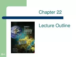
Chapter 14 Lecture Outline. See separate PowerPoint slides for all figures and tables pre-inserted into PowerPoint without notes. 14.1 : Introduction to the Lymphatic System & Immunity. Lymphatic System:

E N D
Chapter 14Lecture Outline See separate PowerPoint slides for all figures and tables pre-inserted into PowerPoint without notes.
14.1: Introduction to the Lymphatic System & Immunity Lymphatic System: • Group of cells and chemicals that travel in lymphatic vessels, and organs & glands that synthesize them • The lymphatic system contains a network of vessels that assists in the circulation of body fluids • Lymphatic vessels collect and carry away excess fluid from interstitial spaces, eventually returning it to the blood • Special vessels called lacteals transport absorbed fats to the circulation • The organs, cells, and biochemicals of the lymphatic system help defend body against disease
14.2: Lymphatic Pathways • Lymphatic pathways start as lymphatic capillaries, that merge to form larger vessels, that empty into veins in the thoracic cavity • Lymphatic Capillaries: • Lymphatic capillaries are tiny, closed-ended tubes that extend into interstitial spaces, paralleling the blood capillaries • Found all over body, except in central nervous system • They receive tissue (interstitial) fluid through their thin walls and slits between cells • Once inside a lymphatic capillary, tissue fluid is called lymph
Lymphatic Vessels • The walls of lymphatic vessels are thinner than those of veins, but are constructed with the same 3 layers • They also have flaplikevalves on the inside, like veins • Larger lymphatic vessels pass through organs called lymph nodes, and then merge to form larger lymphatic trunks
Lymphatic Capillaries Transport Excess FluidBack to the Blood
Valve in a Lymphatic Vessel Insert Figure 14.3 here
Lymphatic Trunks and Collecting Ducts • The lymphatic trunks drain lymph from the lymphatic vessels • The trunks are named for the regions they drain • These trunks empty into 1 of 2 collecting ducts, either the thoracic duct or right lymphatic duct • The right lymphatic duct drains the right side of the head and neck, the right arm, and right thorax, and empties into the right subclavian vein • The thoracic duct, the larger of the collecting ducts,drains the rest (majority) of the body, and empties into the left subclavian vein
14.3: Tissue Fluid and Lymph • Lymph is tissue fluid that has entered a lymphatic capillary; lymph formation depends on tissue fluid formation • Tissue Fluid Formation: • Tissue fluid is made up of water and dissolved substances that leave blood capillaries by filtration and diffusion • Tissue fluid is almost the same as blood plasma, except that it does not contain large plasma proteins; they are too large to pass through capillary walls • Plasma proteins create plasma colloid osmotic pressure, that draws most of the fluid back into the capillaries • Fluid that does not return to the capillaries becomes tissue fluid
Lymph Formation and Function • Filtration from the plasma usually occurs to a greater extent than reabsorption; this leads to tissue fluid formation • Rising osmotic pressure in the tissues interferes with the return of fluids to the bloodstream • Increasing tissue fluid hydrostatic pressure forces some of the fluid into lymphatic capillaries, where it is now called lymph • Most substances, including small proteins, are returned to blood via the lymph • Lymph also transports foreign particles, including bacteria and viruses, to the lymph nodes for recognition and destruction
14.4: Lymph Movement • The hydrostatic pressure of tissue fluid drives the entry of fluid into lymphatic capillaries, where it is now called lymph • Forces that move blood in veins, such as skeletal muscle contraction, breathing movements, contraction of smooth muscle in the walls of the vessels (lymphatic trunks, in this case), and the presence of valves, are also the forces that propel lymph through lymphatic vessels • A condition that interferes with the flow in lymph will result in an accumulation of lymph in the interstitial spaces, called edema • During surgery, lymphatic vessels or tissues may be removed or disturbed, resulting in edema
14.5: Lymphatic Tissues and Organs • Lymphatic tissue contains lymphocytes, macrophages, and other cells • Diffuse, unencapsulated lymphatic tissue associated with the digestive, respiratory, urinary, and reproductive systems is called mucosa-associated lymphoid tissue (MALT) • Tonsils, appendix, and Peyer’s patches are compact masses of lymphatic nodules • Encapsulated lymphatic organs include lymph nodes, the thymus, and spleen
Lymph Nodes • Lymph nodes, which contain lymphocytes and macrophages, are located in groups or chains along lymphatic vessels • Structure of a Lymph node: • Lymph nodes are bean-shaped • Blood vessels, nerves, and efferent lymphatic vessels enter or exit at the indented hilum • Afferent lymphatic vessels enter on the convex surface • Lymph nodes are covered with a connective tissue capsule, that extends inside the node and divides it into nodules and spaces called sinuses • Lymph nodes contain both lymphocytes and macrophages; they filter the lymph as it flows through them, removing many pathogens
Locations of Lymph Nodes • The lymph nodes generally occur in chains along the parts of the larger lymphatic vessels • Lymph nodes are not found in the central nervous system • Major areas of concentrations of lymph nodes: cervical, thoracic, axillary, supratrochlear, abdominal, pelvic, and inguinal regions
Functions of Lymph Nodes Main functions of lymph nodes: • Filter lymph and remove bacteria and cellular debris before lymph is returned to the blood • Immune surveillance: Monitor body fluids; performed by lymphocytes and macrophages • Lymph nodes are also centers of lymphocyte production • Lymphocytes attack viruses, bacteria and parasitic cells that enter a lymph node • Macrophages engulf and destroy foreign particles, debris, and damaged cells
Thymus • The thymus is a soft, bi-lobed organ located behind the sternum, above the heart • It shrinks in size during the lifetime; large in children, small in adults, replaced by adipose & connective tissue in the elderly • The thymus is surrounded by a connective tissue capsule that extends inside it and divides it into lobules • Lobules contain lymphocytes, some of which mature into T cells or T lymphocytes, that leave the thymus to provide immunity • The thymus secretes hormones called thymosins, which influence the maturation of T lymphocytes
Spleen • The spleen lies in the upper left abdominal cavity • Spleen is the body’s largest lymphatic organ • The spleen resembles a large lymph node except that it contains blood instead of lymph • Composed of white pulp, which contains many lymphocytes, and red pulp, which contains red blood cells, macrophages, and lymphocytes • The spleen filters the blood and removes damaged blood cells and bacteria
14.6: Body Defenses Against Infection • Pathogen: A disease-causing agent • Presence and multiplication of a pathogen can produce an infection • Pathogens can be bacteria, viruses, fungi or protozoans • The body has 2 mechanisms of defense against pathogens: • Innate (nonspecific) defenses: • Guard against many types of pathogens; respond quickly • Include species resistance, mechanical barriers, chemical barriers, natural killer cells, inflammation, phagocytosis, and fever • Adaptive (specific) defenses or immunity: • Respond against only a specific type of pathogen; respond more slowly • Accomplished by specialized lymphocytes, which secrete cytokines or antibodies
14.7: Innate (Nonspecific) Defenses Species Resistance: • A species is resistant to diseases that affect other species • Species resistance is based on the following factors: • Different chemical environments in various species • A body temperature that does not provide the conditions required by the pathogens • Presence or absence of receptors for a particular type of pathogen
Mechanical Barriers • Mechanical barriers prevent the entry of certain pathogens by providing a physical separation of pathogens and internal tissues • Examples are the unbroken skin and mucous membranes of the body • Includes hair, mucus, and sweat • Mechanical barriers represent the body’s first line of defense • The rest of the innate defenses are part of the second line of defense
Chemical Barriers • Chemical barriers are chemicals that kill many pathogens • The acidic environment provided by HCl in gastric juice is lethal to some pathogens • Enzymes, such as pepsin in the stomach and lysozyme in tears, destroy many pathogens • Interferons, hormone-like peptides secreted by lymphocytes and fibroblasts when viruses or tumor cells are present, block viral replication and slow the growth of tumors • Complement, a group of proteins in body fluids, stimulates inflammation, attracts phagocytes and enhances phagocytosis
Natural Killer (NK ) Cells • Natural killer (NK) cells are a small group of lymphocytes, other than T cells and B cells • NK cells defend the body against viruses and cancer cells by secreting cytolytic substances called performing, which lyse cell membranes of pathogens • NK cells also secrete chemicals that enhance inflammation
Inflammation • Inflammation is a tissue response to injury or infection • Function of inflammation is to stop the spread of pathogens and infection • Characterized by redness, swelling, heat, and pain • Major actions that occur during an inflammatory response include: • Dilation of blood vessels • Increase of blood volume in affected areas (causes redness) • Invasion of white blood cells into the affected area • Blood clotting & fibrin thread formation • Fibroblasts secrete chemicals that produce a sac around the area to wall off infection; inhibits spread of infection
Phagocytosis • Phagocytosis is the engulfment and digestion of pathogens, foreign particles, and debris • The most active phagocytes are neutrophils and monocytes; these leave the bloodstream by diapedesis (squeezing between adjacent cells of the capillary walls) at areas of injury • They are attracted to the injured area by chemotaxis, which is the process of attraction via chemicals from injured cells • Neutrophils engulf smaller particles; monocytes attack larger ones • Monocytes give rise to macrophages, which are either free or become fixed in various tissues • Neutrophils + monocytes + macrophages = mononuclear phagocytic system or reticuloendothelial system
Fever • A fever occurs when body temperature is re-set to a higher set point by the body, resulting in an elevated body temperature • Fever provides a hostile environment for pathogens that reproduce best under normal human conditions • Elevated body temperature causes the liver and spleen to take up iron, reducing the amount in the blood, and keeping it from fungi or bacteria, which need it for growth and metabolism • Phagocytic cells attack with greater vigor when the temperature rises
14.8: Adaptive (Specific) Defenses or Immunity • Immunity (adaptive immune defenses) is the response by the body against specific pathogens, their toxins, or their metabolic products • This is the body’s third line of defense against pathogens • Performed by lymphocytes and macrophages that recognize and remember specific foreign molecules on particular pathogens
Antigens • Antigens are molecules that that can elicit an immune response • Antigens can be proteins, polysaccharides, glycoproteins, or glycolipids • Before birth, the body makes an inventory of “self” proteins and other large molecules • Immune response is directed against “nonself” molecules, which are usually large and complex foreign molecules • Sometimes small molecules called haptens, which are not antigenic by themselves, combine with larger molecules and become antigenic
Lymphocyte Origins • During fetal development, red bone marrow begins releasing undifferentiated lymphocyte precursors into circulation; this process continues throughout entire lifespan • About half of them reach thymus, and specialize into T lymphocytes or Tcells; these become 70-80% of circulating lymphocytes; some settle in the lymph nodes, spleen, and thoracic duct • Other lymphocytes differentiate in red bone marrow to become Blymphocytes or B cells; these represent 20-30% of circulating lymphocytes; they also settle in the lymph nodes, spleen, and lining of the intestines
Scanning Electron Micrograph of a B Cell Insert Figure 14.12 here
T Cells and the Cellular Immune Response • Lymphocytes require activation before they can respond to antigens • T cell activation requires an encounter with an antigen-presenting cell (accessory cell, APC), such as a B cell or macrophage, that has already encountered and perhaps phagocytized the antigen • Macrophages acting as APCs digest the pathogen, and display the antigenic fragments on their own cell membrane, complexed with special proteins called major histocompatibility complex (MHC) proteins • MHC proteins help T cells recognize displayed antigens • When T cells recognize and bind to antigenic fragments that match their receptors, they become activated
T Cells and the Cellular Immune Response (2) • Activated T cells interact directly with antigen-bearing cells; this type of cell-to-cell contact is called the cellular immune response or cell-mediated immunity • T cells also synthesize and secrete cytokines that enhance cellular responses to antigens • Examples of cytokines are interleukins and colony stimulating factors • Some T cells secrete toxins, growth-inhibiting factors, or interferon
T Cells and the Cellular Immune Response (3) Types of T cells: • Helper T cells stimulate B cells to produce antibodies against the displayed antigen • Cytotoxic Tcells monitor the body's cells, recognizing and eliminating cancer cells and virus-infected cells • Cytokines from helper T cells activate cytotoxic T cells, which then increase number of identical cells in their clone • Cytotoxic T cells then bind to antigen-bearing cells, and release perforin, which cuts pores in the cell membrane, destroying the cells • Memory T cells provide a quick response to any future exposure to the same antigen, by dividing to produce a large number of cytotoxic T cells
B Cells and the Humoral Immune Response • A B cell may become activated and produce a clone of cells when it encounters an antigen that matches its receptors, and binds to it • But most B cells need helper T cells for activation • When a helper T cell encounters a B cell that has already encountered and bound to an antigen, the helper T cell releases cytokines that activate the B cell, stimulating it to proliferate (divide and form a clone) • Some of the B cells differentiate into plasma cells, which produce and secrete antibodies(immunoglobulins) • Antibodies travel through the body fluids to attack and destroy antigens; this is called the humoral immune response • Other B cells become memory B cells; these remain dormant at the time, but respond to future encounters with the antigen
Steps in Antibody Production Insert Table 14.2 here
Types of Antibodies • There are 5 major types of antibodies (immunoglobulins) that constitute the gamma globulin fraction of the plasma • Most abundant are IgG, IgA, and IgM • Types of antibodies: • Immunoglobulin G (IgG) is found in tissue fluid and plasma, and defends against bacterial cells, viruses, and toxins; also activates complement • IgA is found in exocrine gland secretions (breast milk, saliva, tears, nasal fluid, gastric and intestinal juices, bile, urine) • IgM is found in plasma, activates complement, and contains Anti-A and Anti-B, which react with certain red blood cells during transfusions of mismatched blood • IgDis found on the surface of most B lymphocytes, and functions in B cell activation • IgE is found on surfaces of basophils and mast cells; associated with allergic reactions
Antibody Actions 3 methods by which antibodies react to antigens: • Direct attack by agglutination, precipitation, or neutralization of antigens; these methods make antigens more susceptible to phagocytosis • Activation of complement results in opsonization, chemotaxis, inflammation, agglutination, neutralization, alteration, or lysis of antigens or antigen-bearing cells • Inflammation: Stimulation of local inflammatory changes in the area, that helps prevent the spread of the pathogens