Download
1 / 39
390 likes | 566 Views
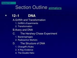
Section 12-1. Section Outline animations. 12–1 DNA A. Griffith and Transformation 1. Griffith’s Experiments 2. Transformation B. Avery and DNA C. The Hershey-Chase Experiment 1. Bacteriophages 2. Radioactive Markers D. The Structure of DNA 1. Chargaff’s Rules 2. X-Ray Evidence

E N D
Section 12-1 Section Outline animations • 12–1 DNA A. Griffith and Transformation 1. Griffith’s Experiments 2. Transformation B. Avery and DNA C. The Hershey-Chase Experiment 1. Bacteriophages 2. Radioactive Markers D. The Structure of DNA 1. Chargaff’s Rules 2. X-Ray Evidence 3. The Double Helix Go to Section:
DNA What is Morse code? --- -- - --- - What is the function? How do organisms pass on traits to offspring? Does every cell of your body contain the same amount of DNA? What about egg and sperm cells? Does replication of DNA need to be accurate? Why or Why not? Why is it important to decode any code?
History of DNA discoveries – (very recent –this century) Griffith ( 1928) - Transformation- one strain of bacteria had been transformed into another. See experiment. Can you imagine producing a tiger from a tabby cat? This was a great breakthrough! Avery and scientists (1944) – What molecules caused this transformation? They discovered DNA was the factor that was an influence on heredity. Hersey and Chase (1952) – Experiments with bacteriophages (bacteria eaters) showed that DNA was the carrier for the genetic code of organisms. What did they do? Rosalind Franklin (1950’s)- Took X-rays of DNA molecules to discover structure. These were used later by Watson and Crick. Watson and Crick _ (1953) – Young scientists who discover double helix shape of DNA. Shared the Nobel Prize in 1962 with Franklin’s assistant (Franklin had died). Their first models of DNA were of rubber and wooden balls with sticks in between. Human Genome Project- What is this?
Figure 12–2 Griffith’s Experiment Section 12-1 Heat-killed, disease-causing bacteria (smooth colonies) Harmless bacteria (rough colonies) Harmless bacteria (rough colonies) Control(no growth) Heat-killed, disease-causing bacteria (smooth colonies) Disease-causing bacteria (smooth colonies) Dies of pneumonia Dies of pneumonia Lives Lives Live, disease-causingbacteria (smooth colonies) Go to Section:
Figure 12–2 Griffith’s Experiment Section 12-1 Heat-killed, disease-causing bacteria (smooth colonies) Harmless bacteria (rough colonies) Harmless bacteria (rough colonies) Control(no growth) Heat-killed, disease-causing bacteria (smooth colonies) Disease-causing bacteria (smooth colonies) Dies of pneumonia Dies of pneumonia Lives Lives Live, disease-causingbacteria (smooth colonies) Go to Section:
Figure 12–4 Hershey-Chase Experiment Section 12-1 Bacteriophage with phosphorus-32 in DNA Phage infectsbacterium Radioactivity inside bacterium Bacteriophage with sulfur-35 in protein coat Phage infectsbacterium No radioactivity inside bacterium Go to Section:
Figure 12–4 Hershey-Chase Experiment Section 12-1 Bacteriophage with phosphorus-32 in DNA Phage infectsbacterium Radioactivity inside bacterium Bacteriophage with sulfur-35 in protein coat Phage infectsbacterium No radioactivity inside bacterium Go to Section:
Figure 12–4 Hershey-Chase Experiment Section 12-1 Bacteriophage with phosphorus-32 in DNA Phage infectsbacterium Radioactivity inside bacterium Bacteriophage with sulfur-35 in protein coat Phage infectsbacterium No radioactivity inside bacterium Go to Section:
What is DNA? -Deoxyribonucleic Acid – Nucleic acid which stores and transmits genetic information. Function of DNA – control of the cell and production of proteins in the cell. *Composed of3 parts: 1. Phosphate group (P) 2. Nitrogen base – 2 types: A. Purines – double ring of carbon and nitrogen (adenine and guanine) guanine adenine B. Pyrimidines – single ring of carbon and nitrogen (thymine and cytosine) cytosine thymine 3. Sugar group *A nucleotide is composed of a nitrogen base, phosphate and sugar. Two complementary nucleotides joined are called a molecule of DNA.
Percentage of Bases in Four Organisms Section 12-1 Source of DNA A T G C Streptococcus 29.8 31.6 20.5 18.0 Yeast 31.3 32.9 18.7 17.1 Herring 27.8 27.5 22.2 22.6 Human 30.9 29.4 19.9 19.8 Go to Section:
Figure 12–5 DNA Nucleotides Purines Pyrimidines Adenine Guanine Cytosine Thymine Deoxyribose Phosphate group Go to Section:
Figure 12–7 Structure of DNA Nucleotide Hydrogen bonds Sugar-phosphate backbone Key Adenine (A) Thymine (T) Cytosine (C) Guanine (G) Go to Section:
Interest Grabber Section 12-2 • A Perfect Copy • When a cell divides, each daughter cell receives a complete set of chromosomes. This means that each new cell has a complete set of the DNA code. Before a cell can divide, the DNA must be copied so that there are two sets ready to be distributed to the new cells. Go to Section:
Section 12-2 Interest Grabber continued 1. On a sheet of paper, draw a curving or zig-zagging line that divides the paper into two halves. Vary the bends in the line as you draw it. Without tracing, copy the line on a second sheet of paper. 2. Hold the papers side by side, and compare the lines. Do they look the same? 3. Now, stack the papers, one on top of the other, and hold the papers up to the light. Are the lines the same? 4. How could you use the original paper to draw exact copies of the line without tracing it? 5. Why is it important that the copies of DNA that are given to new daughter cells be exact copies of the original? Go to Section:
Section Outline Section 12-2 12–2 Chromosomes and DNA Replication A. DNA and Chromosomes 1. DNA Length 2. Chromosome Structure B. DNA Replication 1. Duplicating DNA 2. How Replication Occurs Go to Section:
12-2 DNA Replication – This is how new DNA is made for new cells and for repairing DNA. DNA- must be copied exactly like blueprints. It does this by “UNZIPPING” each side of the double helix. DNA helicase (an enzyme) breaks the hydrogen bonds between nitrogen bases. Polymerase catalyses the new bonds. *DNA can be easily damaged by certain things. What are some of the factors that can damage and change DNA???? The cells have a built in “proofreading” function. This is taken care of by enzymes (which are all proteins), in each cell. The enzymes remove and replace damaged nucleotides to keep the DNA accurate. Accuracy must be maintained since the sequence of nitrogen bases contains the information determining the structure and function of the entire organism –even humans.
Prokaryotic Chromosome Structure E.coli bacterium Chromosome Bases on the chromosome Go to Section:
Figure 12-10 Chromosome Structure of Eukaryotes Nucleosome Chromosome DNA double helix Coils Supercoils Histones Go to Section:
Figure 12–11 DNA Replication Section 12-2 Original strand New strand DNA polymerase Growth DNA polymerase Growth Replication fork Replication fork Nitrogenous bases Original strand New strand
Section Outline Section 12-3 • 12–3 RNA and Protein Synthesis A. The Structure of RNA B. Types of RNA C. Transcription D. RNA Editing E. The Genetic Code F. Translation G. The Roles of RNA and DNA H. Genes and Proteins Go to Section:
Section 12-3 • RNA- Ribonucleic Acid • Function- transmits information for the manufacture proteins. • DNA never leaves the nucleus, but RNA does. • Structure- RNA is made of nucleotide monomers (pieces). • How do RNA and DNA differ? • DNARNA • two strands one strand • deoxyribose sugar ribose sugar • nitrogen base thymine nitrogen base uracil • There are 3 types of RNA that are made in the nucleus and move to the cytoplasm where proteins are made. Go to Section:
There are 3 types of RNA that are made in the nucleus and move to the cytoplasm where proteins are made. 1. Messenger RNA (mRNA) – This is a single, uncoiled strand that transmits information from DNA to be used during protein synthesis (making of proteins). (Ribbon) 2. Transfer RNA (tRNA) – This is a single strand of RNA that is folder back like a “hairpin” and some bases pair with this shape. There are 20 or more varieties to transfer the amino acids. (Cross) 3. Ribosomal RNA (rRNA)- This is a globular form (round and blob-like) of RNA that is part of ribosomes. The true function is not known, but it aids in the making of proteins. (Round)
How is RNA made in the nucleus? RNA is made in the nucleus from a process called Transcription- this is where important information on the DNA molecule is transferred to a “mobile” form that can be moved to the cytoplasm. Protein Synthesis/translation This is how proteins are made in the cytoplasm. How does the cell know which proteins to make? Where does the information come from? How does it get to the cytoplasm???????? Proteins are made of strands called polypeptides. Each polypeptide is composed of many amino acids (aa) attached to each other.
For a certain protein to be made, the amino acids must be in a certain sequence. The genetic code (DNA) is made of nucleotides that are put into a “mobile form” called RNA (in particular mRNA). MRNA is the complimentary strand to each DNA strand. Three nucleotides hooked together are called a codon. (mRNA) Each codon (a-u-g, g-c-c, u-g-a) recognizes a certain amino acid. A chart for amino acids that are recognized by mRNA condons are found in the text. For example a codon for u-u-c codes for the amino acid alanine. The codon a-c-g codes for threonine. These codons are in a special order to code for the order of the amino acids to produce proteins. Translation- see diagrams for steps to help you understand process
Figure 12–14 Transcription Adenine (DNA and RNA) Cystosine (DNA and RNA) Guanine(DNA and RNA) Thymine (DNA only) Uracil (RNA only) RNApolymerase DNA RNA Go to Section:
Section 12-3 Messenger RNA Ribosomal RNA Transfer RNA Bringamino acids toribosome Combine with proteins tRNA mRNA Carry instructions rRNA DNA Ribosome Ribosomes Concept Map RNA can be also called which functions to also called which functions to also called which functions to from to to make up
Figure 12–18 Translation Nucleus Messenger RNA Messenger RNA is transcribed in the nucleus. mRNA Lysine Phenylalanine tRNA Transfer RNA The mRNA then enters the cytoplasm and attaches to a ribosome. Translation begins at AUG, the start codon. Each transfer RNA has an anticodon whose bases are complementary to a codon on the mRNA strand. The ribosome positions the start codon to attract its anticodon, which is part of the tRNA that binds methionine. The ribosome also binds the next codon and its anticodon. Methionine Ribosome Start codon mRNA Go to Section:
Figure 12–18 Translation (continued) The Polypeptide “Assembly Line” The ribosome joins the two amino acids—methionine and phenylalanine—and breaks the bond between methionine and its tRNA. The tRNA floats away, allowing the ribosome to bind to another tRNA. The ribosome moves along the mRNA, binding new tRNA molecules and amino acids. Growing polypeptide chain Ribosome tRNA Lysine tRNA mRNA Completing the Polypeptide The process continues until the ribosome reaches one of the three stop codons. The result is a growing polypeptide chain. mRNA Translation direction Ribosome Go to Section:
Figure 12–17The Genetic Code Go to Section:
Section Outline Section 12-4 • 12–4 Mutations A. Gene Mutations B. Chromosomal Mutations • Determining the Sequence of a Gene • DNA contains the code of instructions for cells. Sometimes, an error occurs when the code is copied. Such errors are called mutations. Go to Section:
Section 12-4 Interest Grabber continued 1. Copy the following information about Protein X: Methionine—Phenylalanine—Tryptophan—Asparagine—Isoleucine—STOP. 2. Use Figure 12–17 on page 303 in your textbook to determine one possible sequence of RNA to code for this information. Write this code below the description of Protein X. Below this, write the DNA code that would produce this RNA sequence. 3. Now, cause a mutation in the gene sequence that you just determined by deleting the fourth base in the DNA sequence. Write this new sequence. 4. Write the new RNA sequence that would be produced. Below that, write the amino acid sequence that would result from this mutation in your gene. Call this Protein Y. 5. Did this single deletion cause much change in your protein? Explain your answer. Go to Section:
Mutations are one of the factors that cause a change in a population. Most are random. Interactions between genes and the environment are extremely important. Mutations-mistakes made in duplicating genetic information.Types:1. Germ mutations- occur in the sex cells2. Somatic mutations - occur in other body cells. These are not inheritable (cancer) Gene Mutations – involve changes in the nucleotides. Types of gene mutations: point mutation – one nucleotide affected Frameshift mutation – deleted or inserted nucleotide Chromosome mutations - when there is a change in the number or structure of chromosomes. 4 Types: deletions, duplications, inversion and translocation (see diagrams)
Gene Regulation *Gene interactions (Recessive vs. Dominant) recessive genes do not produce the enzyme (protein) for a trait to be demonstrated. Gene expression - genes are not activated at the same time or in the same way. When a product of a gene is being made (protein) the gene is said to be expressed. How does a cell know which genes to turn on? Example Caterpillar - What determines the appearance of a type of caterpillar? *Some caterpillars are changed by the food they eat. (Some look like leaves and others like twigs) *Does the food you eat have the same effect on you?
Gene Mutations:Substitution, Insertion, and Deletion Section 12-4 Deletion Insertion Substitution Go to Section:
Figure 12–20 Chromosomal Mutations Section 12-4 Deletion Duplication Inversion Translocation Go to Section:
Section Outline Section 12-5 12–5 Gene Regulation A. Gene Regulation: An Example B. Eukaryotic Gene Regulation C. Regulation and Development Regulation of Protein Synthesis How does the DNA differ in your muscle cell and skin cells? How about a nerve cell and liver cell? Does every cell do the same job? How does each cell control what it does? Go to Section:
Section 12-5 Things which affect genes:Radiation, hormones, temperature, light conditions in cells, other genes, chemicals, nutrition Operon- gene cluster including the following parts: Inducer - starts the activity of the genes. Proteins are produced. Repressor - Stops the activity of the genes. Production of proteins stop. How does this work? Ex: E-coli bacteria only produces lactose-digesting enzymes in the presence of lactose (double milk sugar). There is a lactose operon which causes this to happen in the bacteria. (see diagram)Why doesn’t the bacteria make the enzyme all the time? Different genes are activated (turned on) in different cells. Introns vs. Exons - introns do not code for proteins and are “cut out before transcription. Hox Genes – genes which control organs and tissues development in certain parts of embryos. They determine body plan. Damage to a hox gene can lead to serious structure problems. Go to Section:
Molecular Genetics Chapter 12 Hox Genes • Hox genes are responsible for the general body pattern of most animals.
Typical Gene Structure Section 12-5 Promoter(RNA polymerase binding site) DNA strand Regulatorysites Stoptranscription Start transcription Go to Section: