THE GUSTATORY SYSTEM Description
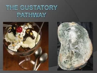
THE GUSTATORY SYSTEM Description The gustatory system is a highly specialized system for reception and processing of the sense of taste. It consists of: The taste buds, which are the peripheral receptors of taste stimuli The gustatory pathway for transmission of gustatory impulse and

THE GUSTATORY SYSTEM Description
E N D
Presentation Transcript
THE GUSTATORY SYSTEM Description The gustatory system is a highly specialized system for reception and processing of the sense of taste. It consists of: The taste buds, which are the peripheral receptors of taste stimuli The gustatory pathway for transmission of gustatory impulse and The cortical centers implicated in gustatory functions.
The Taste Buds Distribution: Taste buds are located in the following areas: • The papillae on the anterior 2/3 of the tongue (Facial nerve). • The posterior 1/3 of the tongue, including vallate papillae (Glosopharyngeal nerve). • The soft palate (Facial nerve). • The pharynx (Glosopharyngeal nerve). • The epiglotis (Vagus nerve).
Component parts of the taste bud: • Gustatory receptor cells: These are ciliated and possess surface receptors for specific flavors and numerous vesicles at the basal aspect of the cells. • Each taste bud receives the branches of the peripheral processes of up to 50 gustatory sensory neurons. (Ratio 1:50) • Types l and ll supportive cells. Both cell types are ciliated but type l possesses apical secretory granules.
Basal cells: These are distributed in the basal part of the taste bud. • They are undifferentiated cells capable of replenishing other cell types of the bud. • von Erbner’s glands: These are serous glands, which secrete a serous fluid that dissolves the tastants. • They are mostly associated with the vallate papillae. • They also secrete lipase, which aids gustation and lipid digestion in the stomach
Taste Pore: This is a pit-like depression in the center of the apical aspect of the bud, which receives glandular secretion required for dissolving materials to be tasted. • The pore is normally filled with a glycoprotein-rich material • Dendritic Synaptic terminals of primary gustatory sensory neurons.
The Gustatory Pathway • This commences with the primary gustatory neurons (1st order neurons) located in three sites viz. • The geniculate ganglion of the facial nerve • The superior and inferior ganglia of the glosopharyngeal nerve and • The inferior (Nodose) ganglion of the vagus nerve.
The peripheral processes of these neurons are distributed to the taste buds in the areas already identified. • The central processes of the three ganglia listed above synapse at gustatory nucleus. in the medulla • Axons of neurons of the gustatory nucleus (2ndorder neurons) project to the thalamus. • The neurons of the gustatory thalamic nucleus (3rd order neurons) project to the primary cortical area for taste, which is located in the postcentralgyrus, adjacent to the first somesthetic area for the tongue. • The primary taste area extends to the upper operculum of the lateral sulcus and to the insula.
THE OLFACTORY SYSTEM This is the highly specialized system adapted for the reception and processing of the sense of smell. It is composed of: • The olfactory Epithelium • The olfactory nerve. • The olfactory bulb. • The olfactory tract. • Cerebral olfactory centers. • Associated olfactory sub-cortical centers.
The Olfactory Epithelium • The olfactory epithelium is a modified pseudostratified ciliated columnar epithelium of the respiratory tract. • It is ectodermal in origin, about 100 micron thick and has a surface area of 10 square centimeters. • It is located in the roof of the nasal cavity extending slightly to medial and lateral walls of the cavity. • It is yellowish in color in the fresh state.
Other salient features of the epithelium are: 1. Olfactory neurosensory cells (Primary olfactory neurons): • These are special bipolar neurons characterized by apical distention (olfactory vesicle) and long, non-motile apical cilia, which are chemoreceptive. • Each nasal cavity may contain as much as 25 million cells, which are continuously replaced at a turn over rate of 3 months. • The axons of these cells form about 20 bundles of unmyelinated olfactory nerves (olfactory fila) from each nasal epithelium. • These nerves traverse the cribriform plate to gain access to the olfactory bulb 2. Pigmented supportive sustentacular cells: These cells possess yellowish pigment, which impacts a yellowish color on the epithelium.
3. Bowman’s glands: These are branched tubular glands, which secrete a lysozyme, and a glycoprotein mucous fluid, which dissolves the odoriferous substance. The mucus contains odorand-binding proteins. 4. Basal cells: These are undifferentiated cells located at the basal aspect of the epithelium and have the capability of replenishing all the cell types in the epithelium. 5. Sensory endings of the trigeminal nerve for perception of noxious sensations
The Olfactory Bulb • This is a bulbous dilation at the commencement of the olfactory tract. • It receives the first order neurons of the olfactory pathway as well as the efferents of the olfactory system. • It is related to the ventral end of the olfactory sulcus of the orbital surface of the frontal lobe. • It contains the second order neurons of the olfactory pathway.
Olfactory Tract • Each olfactory tract consists of myelinated axons principal cells (Mitral cells) of the olfactory bulb and fibres of neurons of the anterior olfactory nucleus • The tract converges on the olfactory trigone located anterior to the anterior perforated substance.
Cortical Olfactory Areas (Centers) There are two primary olfactory areas in the brain,located in the temporal lobe. The two areas are: • The lateral Olfactory Area and • The intermediate Olfactory area • A small area of the insula is also implicated in olfactory function. • An association olfactory area is also present. • This is located in the orbital surface of the frontal lobe
The Olfactory Pathway • The first order neurons of the olfactory pathway are the neurosensory cells of the olfactory epithelium. These unmyelinated fibers form about 20 bundles of olfactory nerve, which traverse the olfactory foramina in the cribriform plate of the ethmoidal bone before entering the olfactory bulb. • The second order neurons are the mitral cells of the olfactory bulb, which give rise to myelinated fibers, which constitute the olfactory tract.
Fibers of this tract converge on the olfactory trigone from where they diverge to pass into the lateral and intermediate olfactory striae. • Lateral olfactory striae fibers project to the lateral olfactory area while • The intermediate striae fibers project to the intermediate olfactory area. • Collaterals of mitral and tufted neurons project to the anterior olfactory nucleus